A Mushroom Rainbow To Put The Fun In Fungi. Cause They Don’t Need Psilocybin to Be Magic. And Though
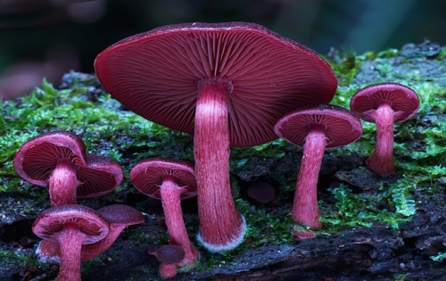
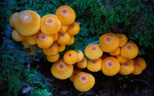
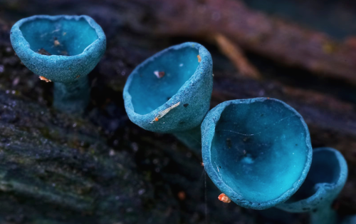
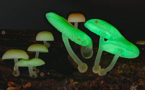

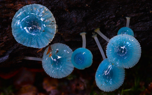
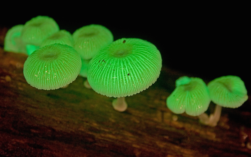
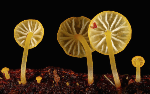
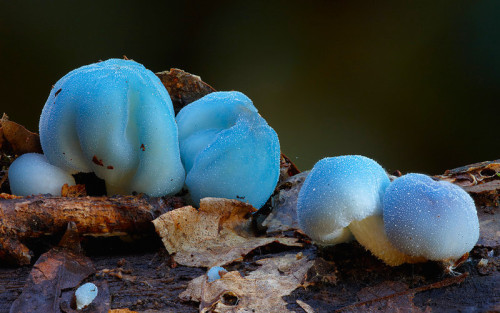
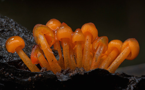
a mushroom rainbow to put the fun in fungi. cause they don’t need psilocybin to be magic. and though some mushrooms are coloured as a toxicity warning to predators, many others are brightly coloured to instead attract potential spore dispersers. see this for more on the bioluminescent mushrooms seen here. (photos)
More Posts from Contradictiontonature and Others
Thyroid function tests

Standard Tests
TSH levels
Free T4 (fT4) levels
Measurements of total T4 + T3 used to be common however detects both bound and free T3 + T4
Elevated total T4 may occur in healthy individuals if there is an increase in binding protein concentrations
Reliable tests now exist for free T4 + T3
T3 = 3.9-6.7 pmol/L
T4 = 12-22 pmol/L
Thyroid-stimulating hormone
Produced by the pituitary gland, not the thyroid, however:
TSH levels are controlled by negative feedback – can be indication of thyroid function (changes in T3+T4 will result in changes in TSH to try compesate)
TSH levels greatly elevated in hypothyroidism – >10 fold increase over reference values
More sensitive marker than decreased fT4 - increased TSH occurs before fT4 decreases
TSH levels greatly supressed in hyperthyroidism
Low concentrations can also occur in non-thyroidal illness
TSH measurement is the first-line test of thyroid function.
Free T4 + T3 Measurements
Desirable as free hormone is clinically relevant
Total levels can change under conditions that alter thyroxine-binding globulin (TBG) levels e.g. pregnancy
Large changes in TBG may still affect fT4 + fT3 levels
fT3 levels often normal in hypothyroidism
fT3 levels usually raised more than fT4 levels in hyperthyroidism
Unless complicated by an illness effecting conversion of T4 to T3
Therefore: – fT4 levels are a better indication of hypothyroidism
fT3 levels are a better indication of hyperthyroidism


Even after someone is declared dead, life continues in the body, suggests a surprising new study with important implications.
Gene expression — when information stored in DNA is converted into instructions for making proteinsor other molecules — actually increases in some cases after death, according to the new paper, which tracked postmortem activity and is published in the journal Open Biology.
“Not all cells are ‘dead’ when an organism dies,” senior author Peter Noble of the University of Washington and Alabama State University told Seeker. “Different cell types have different life spans, generation times and resilience to extreme stress.”
In fact, some cells seem to fight to live after the organism has died.
“It is likely that some cells remain alive and are attempting to repair themselves, specifically stem cells,” Noble said.

Happy Valentine’s Day! Hope everybody gets their share of dopamine and oxytocin today. #lovefeelings #scientificliteracy #braininlove #brainfeels


Scientists are pretty sure that deep inside the moon, there’s water
While Earth’s surface cracks and spouts fire, the moon’s surface, for as long as we’ve known it, has been quiet.
The youngest sign of volcanic activity scientists have found on the moon’s surface is 18 million years old.
But the traces of that long-ago volcanic activity could help scientists crack an enduring mystery: How much water is on the moon?
A study published Monday in Nature Geoscience suggests it may be more than we thought. Read more (7/24/17)
follow @the-future-now

We Just Moved One Step Closer to Understanding (and Defeating) Alzheimer’s


One of the largest icebergs ever recorded, packing about a trillion tons of ice or enough to fill up two Lake Eries, has just split off from Antarctica, in a much anticipated, though not celebrated, calving event.
A section of the Larsen C ice shelf with an area of 2,240 square miles (5,800 square kilometers) finally broke away some time between July 10 and today (July 12), scientists with the U.K.-based MIDAS Project, an Antarctic research group, reported today.
Continue Reading.
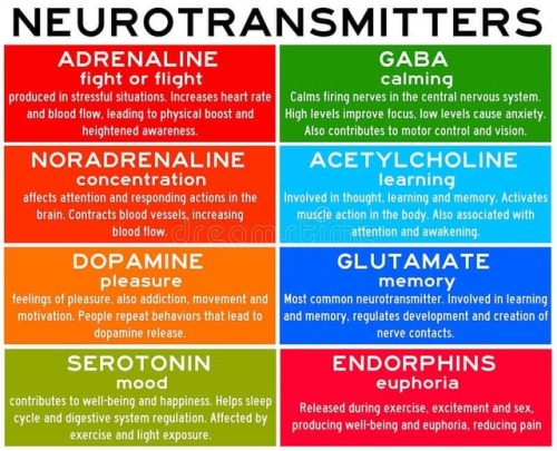
Neurotransmitters are chemicals that help in transmitting signals across a synapse. Different neurotransmitters are associated with different functions. Knowledge about these helps us to treat various neurological conditions by either stimulating or inhibiting these production. #neurology #neuroscience #psychiatry #medicine #medstudynotes #medschool #mbbs #unimed #brain #nervoussystem #physiology #medblog #medblr #medstudent https://www.instagram.com/p/BrM4ocsBqJe/?utm_source=ig_tumblr_share&igshid=12tojib83c32d

A mighty membrane that twists and turns through the gut is starting the new year with a new classification: the structure, called the mesentery, has been upgraded to an organ.
Scientists have known about the structure, which connects a person’s small and large intestines to the abdominal wall and anchors them in place, according to the Mayo Clinic. However, until now, it was thought of as a number of distinct membranes by most scientists. Interestingly, in one of its earliest descriptions, none other than Leonardo da Vinci identified the membranes as a single structure, according to a recent review.

(Image caption: The brain of a fruit fly contains many different regions responsible for processing sight, smell and taste in addition to regions for controlling movement. This image shows the results of a new method which automatically identifies these brain regions. Each color represents a different brain region. The authors used this method to discover specific areas involved in processing of visual information in the fly. The technique could also be used to refine our understanding of vertebrate brains)
Identifying Brain Regions Automatically
Using the example of the fruit fly, a team of biologists led by Prof. Dr. Andrew Straw has identified patterns in the genetic activity of brain cells and taken them as a basis for drawing conclusions about the structure of the brain. The research, published in Current Biology, was conducted at the University of Freiburg and at the Research Institute of Molecular Pathology (IMP) in Vienna, Austria.
The newly developed method focuses on enhancers, DNA segments responsible for enhancing transcription of RNA at specific locations and developmental times in an organism. The research started with a database of three-dimensional images showing individual enhancer activity. The team used an automatic pattern finding algorithm to identify genetic activity patterns shared across the images. They noticed that, in some cases, these patterns seemed to correspond with specific brain regions. To demonstrate the functionality of their method, the biologists began by applying it to regions of the fruit fly brain whose anatomy is already well known – namely, those responsible for the sense of smell. The activity patterns of the enhancers traced the already familiar anatomy of these regions.
Then the biologists used the new method to study brain regions responsible for vision. These experiments led to new insights into the anatomy of these areas: In addition to eleven already known regions, the activity patterns of the enhancers revealed 14 new regions, each of which presumably serves a different function for the fruit fly’s sense of sight. The researchers now aim to conduct further studies to determine which regions are responsible for which functions.
Andrew Straw has served since January 2016 as professor of behavioral neurobiology and animal physiology at the University of Freiburg’s Faculty of Biology and is a member of the Bernstein Center Freiburg (BCF). Before their move to Freiburg, he and his research assistants Karin Panser and Dr. Laszlo Tirian worked at the Research Institute of Molecular Pathology in Vienna in collaboration with Dr. Florian Schulze, Virtual Reality and Visualization Research Center GmbH (VRVis). The goal of Straw’s research is to achieve a better understanding of the structure and function of the brain. He hopes this basic research will ultimately help in the design of therapies for patients suffering from neurological diseases affecting specific regions of the brain.
Results and visualizations: https://strawlab.org/braincode
-
 kryskabs liked this · 8 months ago
 -
 kittylrose liked this · 9 months ago
 -
 endlessbeautifulforms reblogged this · 9 months ago
 -
endlessbeautifulforms liked this · 9 months ago
-
 theuncoolcatapiller liked this · 9 months ago
 -
 soongtypehuman liked this · 9 months ago
 -
 alialink liked this · 9 months ago
 -
 redacted-nameless liked this · 9 months ago
 -
 nists reblogged this · 9 months ago
 -
 maximumcreationcollection liked this · 9 months ago
 -
 sparkling-september reblogged this · 1 year ago
 -
 vinicontraste reblogged this · 1 year ago
 -
vinicontraste liked this · 1 year ago
-
 reeves-and-crows liked this · 1 year ago
 -
 tentoriumcerebelli reblogged this · 1 year ago
 -
 cheese-stands-alone reblogged this · 1 year ago
 -
 hippiesamy reblogged this · 1 year ago
 -
 meta-metamorph reblogged this · 1 year ago
 -
meta-metamorph liked this · 1 year ago
-
tentoriumcerebelli liked this · 1 year ago
-
 kalilaksxlx reblogged this · 1 year ago
 -
 wyrmeleon reblogged this · 1 year ago
 -
 bloody-fate reblogged this · 1 year ago
 -
 anthropomorphicbeancake reblogged this · 1 year ago
 -
bloody-fate liked this · 1 year ago
-
 rainbow-smite reblogged this · 1 year ago
 -
 swtfrmx reblogged this · 1 year ago
 -
 oceanmonument reblogged this · 1 year ago
 -
 bestgore reblogged this · 1 year ago
 -
 troutreznor liked this · 1 year ago
 -
 verminhost reblogged this · 1 year ago
 -
jakeoversee liked this · 1 year ago
-
 mossywombat reblogged this · 1 year ago
 -
 elvn999 reblogged this · 1 year ago
 -
elvn999 liked this · 1 year ago
-
 poppiesandpromises liked this · 1 year ago
 -
 truegrublord reblogged this · 1 year ago
 -
 dapagoldtimow liked this · 1 year ago
 -
 infantisimo liked this · 1 year ago
 -
 dogsandcatsandmiscellaneousstuff reblogged this · 1 year ago
 -
dogsandcatsandmiscellaneousstuff liked this · 1 year ago
-
 petrichornial reblogged this · 1 year ago
 -
 jsttblg reblogged this · 1 year ago
 -
 omaunk reblogged this · 1 year ago
 -
 spice-posh reblogged this · 1 year ago
 -
 andrasta14 reblogged this · 1 year ago
A pharmacist and a little science sideblog. "Knowledge belongs to humanity, and is the torch which illuminates the world." - Louis Pasteur
215 posts