Linda Imagem.
Linda imagem.

A stereo image of Jupiter’s atmosphere, derived from data collected by the Juno spacecraft during perijove 3. These images, taken at different point in the orbit, can be combined to reveal the 3D structure and relief of clouds in the southern atmosphere. To see the image in 3D, relax the eyes until the white circles overlap, then look at the image. Alternatively the image can be viewed with Google Cardboard or another VR device.
Image source: NASA
Processing: James Tyrwhitt-Drake
More Posts from Ritasakano and Others

Ernst Haeckel’s “Paleontological Tree of Vertebrates” (c. 1879).
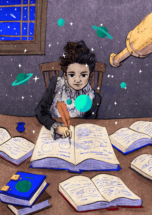
On this day but in 1750, Caroline Lucretia Herschel was born.
Caroline Herschel was the sister of the astronomer William Herschel. After learning astronomy alone and math with the help of her brother, she became his assistant. His most significant contribution to astronomy were the discoveries of various comets, especially comet 35P / Herschel-Rigollet.
🍂 Outono 🍂
Folhas 🍁

via :))) by Inna Dubrovskaya / 500px Autumn Leaves
É lindo quando eles aparecem no meu jardim!!!
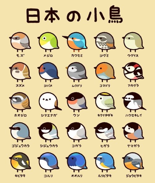
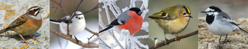
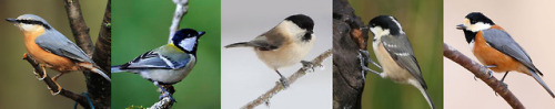
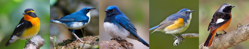
Nihon no kotori (Japanese small birds), cute helpful chart by @T_marohiko listing the following species:
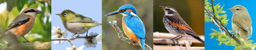
First row - 百舌 mozu (bull-headed shrike) / 目黒 meguro (bonin white-eye) / 川蝉 kawasemi (kingfisher) / ツグミ tsugumi (dusky thrush) / 鶯 uguisu (japanese bush warbler)
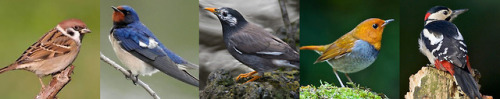
Second row - 雀 suzume (sparrow) / 燕 tsubame (swallow) / 椋鳥 mukudori (grey starling) / 駒鳥 komadori (japanese robin) / 赤啄木鳥 akagera (great spotted woodpecker)
Third Row - 頬白 hoojiro (meadow bunting) / シマエナガ shimaenaga (silver-throated dasher) / 鷽 uso (japanese bullfinch) / 菊戴 kikuitadaki (goldcrest) / 白鶺鴒 hakusekirei (black-backed wagtail)
Fourth row - 五十雀 gojuukara (eurasian nuthatch) / 四十雀 shijuukara (japanese tit) / 小雀 kogara (willow tit) / 日雀 higara (coal tit) / 山雀 yamagara (varied tit)
Fifth row - 黄鶲 kibitaki (narcissus flycatcher) / 小瑠璃 koruri (siberian blue robin) / 大瑠璃 ooruri (blue-and-white flycatcher) / 瑠璃鶲 ruribitaki (red-flanked bluetail) / 尉鶲 joubitaki (daurian redstart)

“The Stella Pinafore Toilette.”
Enquire Within, Ladies Home Journal
5th February 1916.

Niigata Vista do Hotel Nikko
Colcha de retalho
Início sempre tem seus cortes tortos, mesmo que se use régua e cortador, parece que a mão gosta de dar outro contorno aos cortes.
Colcha de retalho é cheia de lembranças, conta histórias perdidas no tempo e deixa no presente a satisfação de fazer, para o futuro a certeza que a história continua.


Lindas imagens!
The Beautiful Things inside Your Head: Winners of the 10th Annual Art of Neuroscience Contest
Winner: Lidija Kononenko

Artist Kononenko described this interactive piece as “a microscope specimen, a map of symptoms, and an investigation of the unknown” in a statement accompanying it. Viewers can zoom in and explore the details of a microscope image of the peripheral nerve system, which is overlaid by textual facts and poetic phrases about sleep.Sleep is “a voluntary act of losing one’s own consciousness,” Konenenko explained in her statement. The poetic snippets resemble the fragmented thoughts humans have while falling asleep. And zooming in and out of the image represents the transition between wakefulness and sleep. Additionally, 31-3594 allows the viewer to act as a pathologist, achieving the goal of blending neuroscience and art. In assessing this unique piece, the jurors praised it for “the interactivity and playful combination of imagery of a human peripheral nerve with a text-based story that unfolds at various scales and highlights the role of the nervous system in the human condition.”
Honorable Mentions

Red Haze by Nicki Coveña
A tsunami of red dots dominates this image by neuroscientist Coveña. The bright red color comes from a fluorescent protein, which was used to visualize the workings of TBR1—a gene that synthesizes the protein that regulates the information transfer from DNA to messenger RNA in vertebrate embryo development. “The out-of-focus view makes one guess at what details are hidden below,” the jurors wrote.

Motor White Matter Networks of the Human Brain by Sanja Budisavljevic
In this piece, neuroscientist Budisavljevic superimposed color onto a 19th-century black-and-white drawing of a brain based on a postmortem dissection. Each color indicates a different “highway,” or white matter pathway connecting particular regions of gray matter and allowing information to be transferred. Red indicates the most prominent highway, which links the cortex and spinal cord. “This pathway carries the messages to and from the body and allows us to function in our sensory world,” Budisavljevic says. Green represents the connection that supports coordination, and blue shows the one that regulates movements.

Bdl by Paméla Simard (Alex Tran, photographs)
Artist Simard partnered with Hunter Shaw, a neuroscientist then at McGill University, to create a series of delicate wooden sculptures. “The various installations were created from fluorescent microscopy images representing the visual system of the fruit fly brain,” Simard wrote in her statement. The intricate details of the fruit fly visual system were made possible by first laminating the thin slices of different types of wood together, then hand cutting the result to mimic the microscope images.
More Art from 2020 Gallery

Whale Retina Rainbow by Elena Vecino Cordero and Luis López Vecino
In February 2019 the death of a whale in Sopelana Beach in Spain made the local news. The beach happened to be close to the University of the Basque Country, where biologist Vecino Cordero works. Seizing the opportunity, she and some volunteers extracted the eye of the whale and took it back to her ophthalmology research group for further study. The image was produced as a part of their research. The whale’s retina was imaged using scanning electron microscopy. And later López Vecino added the colors using Adobe Photoshop.

Sensing Spin by Dan Jagger
Physiologist Jagger used a high-resolution microscope to capture this image. It shows mechanosensory hair cells located in the inner ear that play a role in the sense of balance. A protein called actin is within bundles of stereocilia and is stained yellow. Actin helps the bundles to stand upright, so when the human head turns, they can detect the movement of the fluid they are immersed in. The hair-cell nuclei are stained with cyan.

The Protection of Nature Starts in Our Mind by Robert Luck
Luck is a neuroscientist at Heidelberg University in Germany who studies the development of the cerebellum, located where the spinal cord meets the brain. Alarmed by climate change and deforestation, he created a “mind forest” that resembles bird’s-eye-view photographs of real forests. The “trees” are 65 individually traced images of mice’s Purkinje neurons, which play important roles in controlling coordination and movements. “I chose the number 65 to represent the number of years needed for the rainforest to regrow and gain back at least 80% of its diversity,” Luck wrote in his statement. “[Sixty-five] years—a human lifetime!”

Memories and Patterns: Oligodendrocytes by Shanthi Chandrasekar
Oligodendrocytes are glial cells that support and insulate long neuronal axons. The cells’ lipid membrane wraps around the axons to strengthen the structure, as well as to help neurons to send signals quickly. “A single oligodendrocyte can connect with multiple axons,” artist Chandrasekar wrote in her statement. “In this [pen-and-ink] drawing, I have tried to bring out the connectedness of the oligodendrocytes and the axons.”

Shelter in Place by Geinene Carson
As its title suggests, this piece represents “the artist’s interpretation of the pandemic experience” while sheltering in place because of COVID-19, according to artist Carson’s statement. This acrylic-on-canvas piece is a part of a series entitled Neuron, which started as “visual prayers for our daughter with a rare genetic disorder,” Carson wrote on her Web site. While Shelter in Place implies physical restrictions, Carson, who is based in Atlanta, draws inspiration from the neural network, “because as important as our physical surroundings are to our state of living, our thought life holds the key to thriving within whatever the circumstances may be,” she wrote.

Bridges between Genesis and Neuroscience: Triplets by Rui Rodrigues
This image features three neurospheres—clusters of neural stem or progenitor cells—that are similar in size and shape. Because of their similarity, neurobiologist Rodrigues entitled the piece Triplets. The vibrant colors come from “antibodies coupled with fluorescent tags to label specific proteins,” he says.

The Transfer by Geinene Carson

Motor Neuron Architectural Digest by Stefanie Hauck - University of Bonn

Illuminating The Vascular Network - EPFL by Marwan Abdellah
Whale automata by Sylvain Gautier.

-
 thatforestprince liked this · 1 year ago
 -
 furiouscreatorempathcloud liked this · 7 years ago
 -
 felunax liked this · 7 years ago
 -
 starlovely liked this · 8 years ago
 -
 ritasakano reblogged this · 8 years ago
 -
ritasakano liked this · 8 years ago
-
 tthomusic liked this · 8 years ago
 -
 orions-underpants liked this · 8 years ago
 -
 oldevil liked this · 8 years ago
 -
tenderlysteadycoffee-blog liked this · 8 years ago
-
 gangnam-opevent-blog reblogged this · 8 years ago
 -
 cambodiaeducationfund-blog1 liked this · 8 years ago
 -
 survivingtherenaissance reblogged this · 8 years ago
 -
 downtherabbitholeee reblogged this · 8 years ago
 -
 littleplasticspaceship reblogged this · 8 years ago
 -
 salemsrealm reblogged this · 8 years ago
 -
 bitchydefendorcoffee-blog liked this · 8 years ago
 -
 arazoa reblogged this · 8 years ago
 -
 jmpauly liked this · 8 years ago
 -
 sinnephi liked this · 8 years ago
 -
halloholograms liked this · 8 years ago
-
 tsuchiman reblogged this · 8 years ago
 -
tsuchiman liked this · 8 years ago
-
 autumndude liked this · 8 years ago
 -
 nyogut-blog liked this · 8 years ago
 -
 lopaseo liked this · 8 years ago
 -
 lancia1208 reblogged this · 8 years ago
 -
lancia1208 liked this · 8 years ago
-
 tatzelwurming liked this · 8 years ago
 -
 the-nuclear-chaos reblogged this · 8 years ago
 -
 o-blivia reblogged this · 8 years ago
 -
 outreading-blog reblogged this · 8 years ago
 -
 azureberries liked this · 8 years ago
 -
 gregorystees reblogged this · 8 years ago
 -
 jaguar-thornproof reblogged this · 8 years ago
 -
jaguar-thornproof liked this · 8 years ago
-
 atomiconstruct reblogged this · 8 years ago
 -
survivingtherenaissance liked this · 8 years ago