Scalp Is The Soft Tissue Layer Covering The Bony Vault Over The Brain. It Is Usually Described As Having

Scalp is the soft tissue layer covering the bony vault over the brain. It is usually described as having five layers: S: The skin on the head from which head hair grows. It contains numerous sabaeceous glands and hair follicles C: Connective tissue. A thin layer of fat and fibrous tissue lies beneath the skin. A: The aponeurosis called epicranial aponeurosis (or galea aponeurotica) is the next layer. It is a tough layer of dense fibrous tissue which runs from the frontalis muscle anteriorly to the occipitalis posteriorly. L: The loose areolar connective tissue layer provides an easy plane of separation between the upper three layers and the pericranium. P: The pericranium is the periosteum of the skull bones and provides nutrition to the bone and the capacity for repair.
(x)
More Posts from Science-is-magical and Others

From vision to hand action
Our hands are highly developed grasping organs that are in continuous use. Long before we stir our first cup of coffee in the morning, our hands have executed a multitude of grasps. Directing a pen between our thumb and index finger over a piece of paper with absolute precision appears as easy as catching a ball or operating a doorknob. The neuroscientists Stefan Schaffelhofer and Hansjörg Scherberger of the German Primate Center (DPZ) have studied how the brain controls the different grasping movements. In their research with rhesus macaques, it was found that the three brain areas AIP, F5 and M1 that are responsible for planning and executing hand movements, perform different tasks within their neural network. The AIP area is mainly responsible for processing visual features of objects, such as their size and shape. This optical information is translated into motor commands in the F5 area. The M1 area is ultimately responsible for turning this motor commands into actions. The results of the study contribute to the development of neuroprosthetics that should help paralyzed patients to regain their hand functions (eLife, 2016).
The three brain areas AIP, F5 and M1 lay in the cerebral cortex and form a neural network responsible for translating visual properties of an object into a corresponding hand movement. Until now, the details of how this “visuomotor transformation” are performed have been unclear. During the course of his PhD thesis at the German Primate Center, neuroscientist Stefan Schaffelhofer intensively studied the neural mechanisms that control grasping movements. “We wanted to find out how and where visual information about grasped objects, for example their shape or size, and motor characteristics of the hand, like the strength and type of a grip, are processed in the different grasp-related areas of the brain”, says Schaffelhofer.
For this, two rhesus macaques were trained to repeatedly grasp 50 different objects. At the same time, the activity of hundreds of nerve cells was measured with so-called microelectrode arrays. In order to compare the applied grip types with the neural signals, the monkeys wore an electromagnetic data glove that recorded all the finger and hand movements. The experimental setup was designed to individually observe the phases of the visuomotor transformation in the brain, namely the processing of visual object properties, the motion planning and execution. For this, the scientists developed a delayed grasping task. In order for the monkey to see the object, it was briefly lit before the start of the grasping movement. The subsequent movement took place in the dark with a short delay. In this way, visual and motor signals of neurons could be examined separately.
The results show that the AIP area is primarily responsible for the processing of visual object features. “The neurons mainly respond to the three-dimensional shape of different objects”, says Stefan Schaffelhofer. “Due to the different activity of the neurons, we could precisely distinguish as to whether the monkeys had seen a sphere, cube or cylinder. Even abstract object shapes could be differentiated based on the observed cell activity.”
In contrast to AIP, area F5 and M1 did not represent object geometries, but the corresponding hand configurations used to grasp the objects. The information of F5 and M1 neurons indicated a strong resemblance to the hand movements recorded with the data glove. “In our study we were able to show where and how visual properties of objects are converted into corresponding movement commands”, says Stefan Schaffelhofer. “In this process, the F5 area plays a central role in visuomotor transformation. Its neurons receive direct visual object information from AIP and can translate the signals into motor plans that are then executed in M1. Thus, area F5 has contact to both, the visual and motor part of the brain.”
Knowledge of how to control grasp movements is essential for the development of neuronal hand prosthetics. “In paraplegic patients, the connection between the brain and limbs is no longer functional. Neural interfaces can replace this functionality”, says Hansjörg Scherberger, head of the Neurobiology Laboratory at the DPZ. “They can read the motor signals in the brain and use them for prosthetic control. In order to program these interfaces properly, it is crucial to know how and where our brain controls the grasping movements”. The findings of this study will facilitate to new neuroprosthetic applications that can selectively process the areas’ individual information in order to improve their usability and accuracy.


Olinguito
On Tuesday, Bill Stanley grabbed my arm and pulled me into a side hallway as we were walking towards the mammal collections on the third floor. He looked around suspiciously before leaning in, and in a hushed tone he said
there’s a new raccoon.
What do you mean?
There’s a new raccoon. You can’t tell anyone. It’s in the pipeline. Going live on Thursday.
Wha- I wasn’t going to-
You can’t tell anyone. Guess where it was discovered?
Oh, geez. I don’t know. Maybe Per-
Here. It was discovered here.
Then he patted my shoulder, winked, and kept walking.
Such was my introduction to the olinguito - and yesterday Bill brought it out to show me. In front of us were two drawers, one with the previously known species and the newly described animals on the right. It was immediately obvious to me that before us were two different animals - the size, color and length of the fur, the size of the ears - but without the previous knowledge that they were not one in the same, would I have seen the same dissimilarities?
This is what sparks me. This is what drives my enthusiasm. In these drawers for sixty years, side-by-side these animals remained, their full potential not realized until a curious researcher took the quiet time to sit down and take a concentrated look at them. It’s the romance of the discovery – it was here all along! – and once you see the striking inconsistencies there comes a feeling of empowerment, the thought that we are the next big discoverers. The thrill of the breakthrough remains attainable, accessible. It’s not beyond our reach or out of grasp - I look forward to seeing what you find next.


It’s #InternationalKissingDay! Here’s some topical lipstick chemistry. More info/high-res image: http://wp.me/s4aPLT-lipstick

The Neuroscience of Drumming

According to new neuroscience research, rhythm is rooted in innate functions of the brain, mind, and consciousness. As human beings, we are innately rhythmic. Our relationship with rhythm begins in the womb. At twenty two days, a single (human embryo) cell jolts to life. This first beat awakens nearby cells and incredibly they all begin to beat in perfect unison. These beating cells divide and become our heart. This desire to beat in unison seemingly fuels our entire lives. Studies show that, regardless of musical training, we are innately able to perceive and recall elements of beat and rhythm.
It makes sense then that beat and rhythm are an important aspect in music therapy. Our brains are hard-wired to be able to entrain to a beat. Entrainment occurs when two or more frequencies come into step or in phase with each other. If you are walking down a street and you hear a song, you instinctively begin to step in sync to the beat of the song. This is actually an important area of current music therapy research. Our brain enables our motor system to naturally entrain to a rhythmic beat, allowing music therapists to target rehabilitating movements. Rhythm is a powerful gateway to well-being.
Neurologic Drum Therapy
Neuroscience research has demonstrated the therapeutic effects of rhythmic drumming. The reason rhythm is such a powerful tool is that it permeates the entire brain. Vision for example is in one part of the brain, speech another, but drumming accesses the whole brain. The sound of drumming generates dynamic neuronal connections in all parts of the brain even where there is significant damage or impairment such as in Attention Deficit Disorder (ADD). According to Michael Thaut, director of Colorado State University’s Center for Biomedical Research in Music, “Rhythmic cues can help retrain the brain after a stroke or other neurological impairment, as with Parkinson’s patients ….” The more connections that can be made within the brain, the more integrated our experiences become.
Studies indicate that drumming produces deeper self-awareness by inducing synchronous brain activity. The physical transmission of rhythmic energy to the brain synchronizes the two cerebral hemispheres. When the logical left hemisphere and the intuitive right hemisphere begin to pulsate in harmony, the inner guidance of intuitive knowing can then flow unimpeded into conscious awareness. The ability to access unconscious information through symbols and imagery facilitates psychological integration and a reintegration of self.
In his book, Shamanism: The Neural Ecology of Consciousness and Healing, Michael Winkelman reports that drumming also synchronizes the frontal and lower areas of the brain, integrating nonverbal information from lower brain structures into the frontal cortex, producing “feelings of insight, understanding, integration, certainty, conviction, and truth, which surpass ordinary understandings and tend to persist long after the experience, often providing foundational insights for religious and cultural traditions.”
It requires abstract thinking and the interconnection between symbols, concepts, and emotions to process unconscious information. The human adaptation to translate an inner experience into meaningful narrative is uniquely exploited by drumming. Rhythmic drumming targets memory, perception, and the complex emotions associated with symbols and concepts: the principal functions humans rely on to formulate belief. Because of this exploit, the result of the synchronous brain activity in humans is the spontaneous generation of meaningful information which is imprinted into memory. Drumming is an effective method for integrating subjective experience into both physical space and the cultural group.

ELI5:How come people can't be cryogenically frozen safely as the ice crystals destroy the cell membranes, but sex cells such as sperm are kept frozen for long periods of time yet remain functional?
I work in a lab where we freeze down cells all of the time. We freeze our cells in a medium that contains 5% DMSO, which among other things can be used as a cryoprotectant. However, DMSO is also toxic to cells at the concentrations necessary for cryoprotection. Consequently, when you freeze cells in DMSO, you add the DMSO medium at ice-cold temperatures and don’t allow the cells to warm up. When you later thaw the cells, you have to dilute out the DMSO as quickly as possible without causing osmotic shock, which can pop the cells. Such restrictions on freezing and thawing would basically be impossible to control at the level of a complete organism.
However, to contradict a lot of previous posts, individual cells can be recovered from freezing with high viability. When performed properly (and this varies quite a bit by cell type), you can expect >90% of cells to be alive following thaw.
The chemicals that allow cells to survive freezing are toxic to the body. Keeping the cells cold minimized the damage that this chemical does to the cells. With single cell solutions, adding the chemical at ice-cold temperatures and immediately diluting it out when you thaw the cells can keep 90% of the cells alive. There’s no way to do this with an intact body.
It’s also worth noting that this is probably not the only reason that this technique doesn’t scale to organisms.
Explain Like I`m Five: good questions, best answers.


It always creeps me out...
…that no matter
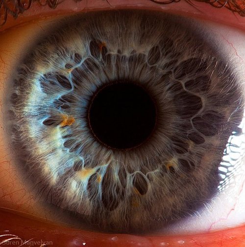
how close
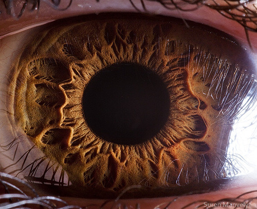
you get
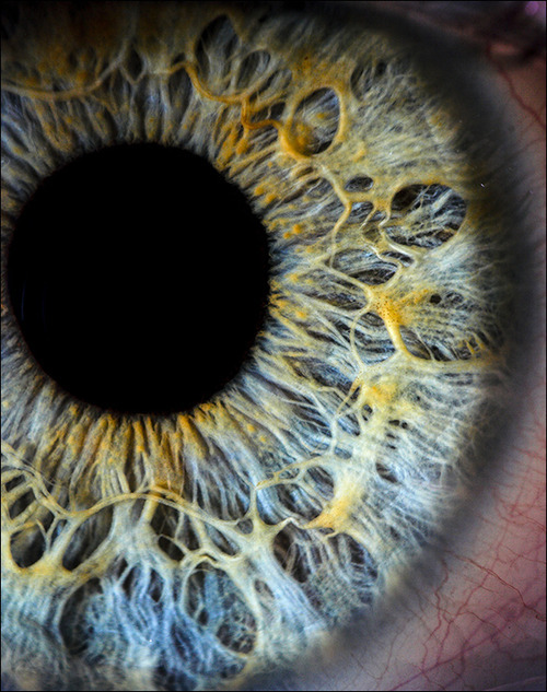
the pupil
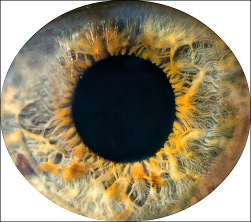
seems to
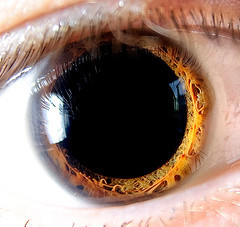
devour light
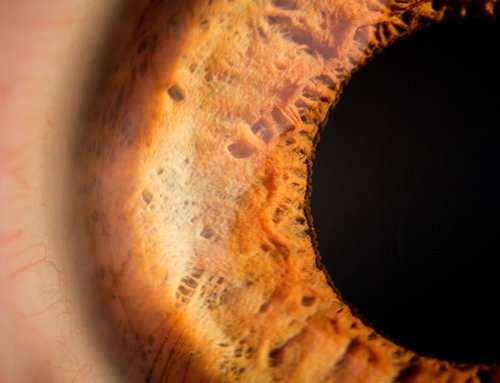
like a black hole
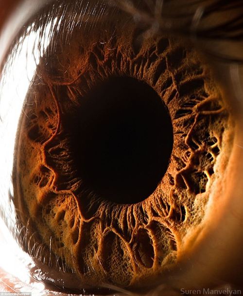
it reflects no light
-
 physistuff liked this · 2 years ago
 -
 distinctlyferal liked this · 4 years ago
 -
 misfit-roo reblogged this · 4 years ago
 -
 graveyardwormss liked this · 5 years ago
 -
 23papercuts liked this · 6 years ago
 -
 alilbitduygu liked this · 7 years ago
 -
 brucewombs liked this · 7 years ago
 -
 ryleegein liked this · 7 years ago
 -
 foroneflower reblogged this · 7 years ago
 -
 biebieee liked this · 7 years ago
 -
 spanish-languagefilm liked this · 7 years ago
 -
 mattwardpictures liked this · 7 years ago
 -
 blackholeson liked this · 7 years ago
 -
 mia-christ-blog liked this · 7 years ago
 -
 brainyey liked this · 7 years ago
 -
 thecorpsecat liked this · 7 years ago
 -
 total-flake reblogged this · 8 years ago
 -
total-flake liked this · 8 years ago
-
 l-a-mazona reblogged this · 8 years ago
 -
 15beans-blog reblogged this · 8 years ago
 -
 bringmethemermaids liked this · 8 years ago
 -
science-is-magical reblogged this · 8 years ago
-
 skyhighrom reblogged this · 8 years ago
 -
skyhighrom liked this · 8 years ago
-
 i-am-the-gatekeeper reblogged this · 8 years ago
 -
 study-md-blog liked this · 8 years ago
 -
 camsterdam-doodles liked this · 8 years ago
 -
 scotty73 liked this · 8 years ago
 -
 the-alphaa liked this · 8 years ago
 -
 catgirlwerewolf liked this · 8 years ago
 -
 shibaka-inu liked this · 8 years ago
 -
 le-piaf reblogged this · 8 years ago
