Science-is-magical - Science Is Magic


More Posts from Science-is-magical and Others

Newborn baby brain scans will help scientists track brain development
Scientists have published ground-breaking scans of newborn babies’ brains which researchers from all over the world can download and use to study how the human brain develops.

(Image caption: Diffusion MRI of the developing neonatal brain (Left: Multi-shell high angular resolution diffusion data decomposed into a free water component (greyscale background image) and a directionally resolved brain tissue component shown as rendered surfaces. Middle and right: Visualisation of anatomical connections in the developing brain derived from the brain tissue component.)
The images are part of the Developing Human Connectome Project (dHCP), a collaboration between King’s College London, Imperial College London and the University of Oxford, which will uncover how the brain develops, including the wiring and function of the brain during pregnancy and how this changes after birth. The dHCP researchers are sharing their images and methods online so that other scientists from around the world can use the data in their own research.
Using Magnetic Resonance Imaging (MRI) scanners at Evelina London Children’s Hospital, the team has developed new techniques which enable images of the brains of foetuses and babies to be captured. Researchers have overcome problems caused by babies’ movement and small size, as well as the difficulties in keeping vulnerable infants safe in the MRI scanner, so that they can now produce highly detailed and rich information on brain development.
The project will help scientists to understand how conditions such as autism develop, or how problems in pregnancy affect brain growth.
‘The Developing Human Connectome Project is a major advance in understanding human brain development - it will provide the first map of how the brain’s connections develop, and how this goes wrong in disease,’ said Lead Principal Investigator, Professor David Edwards from King’s College London and Consultant Neonatologist at Evelina London.
The research collaboration is funded by a €15 million Synergy Grant from the European Research Council, and one of the goals of the project is to make sure that the data is shared as widely across the world as possible. Scientists are able to download the images at https://data.developingconnectome.org.
For this project, Professor Jo Hajnal’s team at King’s College London developed new MRI technology specifically designed to provide high resolution scans of newborn and fetal brains.
In addition, a group led by Professor Daniel Rueckert’s at Imperial College London developed new computer programs to analyse the images. 'We have been developing novel approaches that help researchers by automatically analysing the rich and comprehensive MR images that are collected as part of dHCP,’ he explained.
At the University of Oxford, Professor Steve Smith’s team has been developing specific techniques to define where the connections are in the developing brain.
As well as studying more babies, the team at Evelina London are now recruiting pregnant mothers for foetal scanning. Meanwhile, the first release of images today will allow scientists to start to explore these powerful images and begin mapping out the complexities of human brain development in a whole new way.

From vision to hand action
Our hands are highly developed grasping organs that are in continuous use. Long before we stir our first cup of coffee in the morning, our hands have executed a multitude of grasps. Directing a pen between our thumb and index finger over a piece of paper with absolute precision appears as easy as catching a ball or operating a doorknob. The neuroscientists Stefan Schaffelhofer and Hansjörg Scherberger of the German Primate Center (DPZ) have studied how the brain controls the different grasping movements. In their research with rhesus macaques, it was found that the three brain areas AIP, F5 and M1 that are responsible for planning and executing hand movements, perform different tasks within their neural network. The AIP area is mainly responsible for processing visual features of objects, such as their size and shape. This optical information is translated into motor commands in the F5 area. The M1 area is ultimately responsible for turning this motor commands into actions. The results of the study contribute to the development of neuroprosthetics that should help paralyzed patients to regain their hand functions (eLife, 2016).
The three brain areas AIP, F5 and M1 lay in the cerebral cortex and form a neural network responsible for translating visual properties of an object into a corresponding hand movement. Until now, the details of how this “visuomotor transformation” are performed have been unclear. During the course of his PhD thesis at the German Primate Center, neuroscientist Stefan Schaffelhofer intensively studied the neural mechanisms that control grasping movements. “We wanted to find out how and where visual information about grasped objects, for example their shape or size, and motor characteristics of the hand, like the strength and type of a grip, are processed in the different grasp-related areas of the brain”, says Schaffelhofer.
For this, two rhesus macaques were trained to repeatedly grasp 50 different objects. At the same time, the activity of hundreds of nerve cells was measured with so-called microelectrode arrays. In order to compare the applied grip types with the neural signals, the monkeys wore an electromagnetic data glove that recorded all the finger and hand movements. The experimental setup was designed to individually observe the phases of the visuomotor transformation in the brain, namely the processing of visual object properties, the motion planning and execution. For this, the scientists developed a delayed grasping task. In order for the monkey to see the object, it was briefly lit before the start of the grasping movement. The subsequent movement took place in the dark with a short delay. In this way, visual and motor signals of neurons could be examined separately.
The results show that the AIP area is primarily responsible for the processing of visual object features. “The neurons mainly respond to the three-dimensional shape of different objects”, says Stefan Schaffelhofer. “Due to the different activity of the neurons, we could precisely distinguish as to whether the monkeys had seen a sphere, cube or cylinder. Even abstract object shapes could be differentiated based on the observed cell activity.”
In contrast to AIP, area F5 and M1 did not represent object geometries, but the corresponding hand configurations used to grasp the objects. The information of F5 and M1 neurons indicated a strong resemblance to the hand movements recorded with the data glove. “In our study we were able to show where and how visual properties of objects are converted into corresponding movement commands”, says Stefan Schaffelhofer. “In this process, the F5 area plays a central role in visuomotor transformation. Its neurons receive direct visual object information from AIP and can translate the signals into motor plans that are then executed in M1. Thus, area F5 has contact to both, the visual and motor part of the brain.”
Knowledge of how to control grasp movements is essential for the development of neuronal hand prosthetics. “In paraplegic patients, the connection between the brain and limbs is no longer functional. Neural interfaces can replace this functionality”, says Hansjörg Scherberger, head of the Neurobiology Laboratory at the DPZ. “They can read the motor signals in the brain and use them for prosthetic control. In order to program these interfaces properly, it is crucial to know how and where our brain controls the grasping movements”. The findings of this study will facilitate to new neuroprosthetic applications that can selectively process the areas’ individual information in order to improve their usability and accuracy.
Why we need GMOs to survive climate change
Genetically modified organisms get a bad rap for many reasons, but we’ve actually been genetically altering what we eat since the dawn of human history.
“For 10,000 years, we have altered the genetic makeup of our crops,”explains UC Davis plant pathology professor Pamela Ronald.
“Today virtually everything we eat is produced from seeds that we have genetically altered in one way or another.” (You can read more about Ronald’s thoughts on genetically engineered food here.)
Right now her focus is on rice. It’s one of our basic crops and without it, we would struggle to feed much of the world.
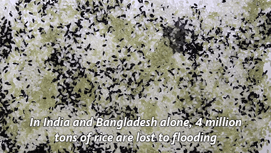
With climate change, we’re seeing an increase in flooding in places like India and Bangladesh, which makes it harder to grow this important food staple.
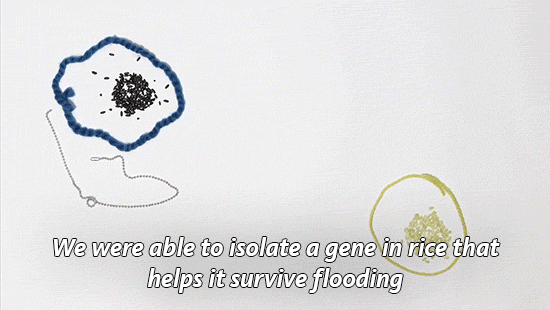
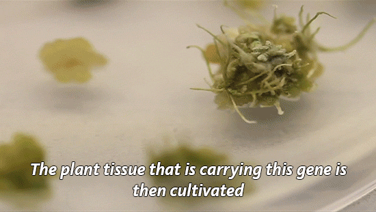
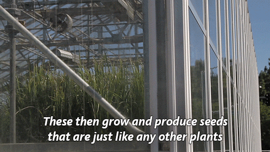
So Ronald and her lab have developed a flood-tolerant strain of rice. It’s known as Sub1a or “scuba rice” and millions of farmers in South Asia are now growing it in their fields.

Today is National Food Day, a day dedicated to hunger awareness. But as we focus on food insecurity, we need to talk more about how global warming will make the problem worse.
As our climate continues to heat up, it has huge impacts on what foods we are able to grow. Will our crops be able to survive droughts and floods? The University of California leads six labs that are working to develop other climate-resilient crops including chickpea, cowpea and millet.
Find out what other scientists are doing to improve our food.
A reminder that NASA isn’t the only space agency
I have seen many “Space achievements 2015” articles and posts leaving international accomplisments completely out, so here are some of them:
1. A new type of basaltic rock on the moon was found by Chinese robotic lander.
China National Space Administration’s Chang’e-3 landed on the Moon on 14 December 2013, becoming the first spacecraft to soft-land since the Soviet Union‘s Luna 24 in 1976.

2. On February 11, the European Space Agency, ESA, successfully launched on a suborbital trajectory and recovered an experimental wingless glider, IXV.
It became the first true “lifting body” vehicle, which reached a near-orbital speed and then returned back to Earth without any help from wings.

3. On December 9, Japan’s Akatsuki spacecraft succeeded entering orbit of Venus.
Japan Aerospace eXploration Agency’s Akatsuki is the first spacecraft to explore Venus since the ESA’s Venus Express reached the end of its mission in 2014.

4. ESA’s Rosetta spacecraft detected oxygen ‘leaking’ from comet 67P/Churyumov-Gerasimenko, the first time these molecules have been seen around a comet.
Rosetta spacecraft, the first to drop a lander (named Philae) on a comet, entered orbit around 67P in 2014 and continues to orbit the body. On June 13, European Space Operations Centre in Darmstadt, Germany, received signals from the Philae lander after months of silence.

5. The Canadian Space Agency has provided NASA with a laser mapping system that will scan an asteroid that could potentially hit the Earth in about 200 years

6. The high-resolution stereo camera on ESA’s Mars Express captured this sweeping view from the planet’s south polar ice cap and across its cratered highlands and beyond.

find Sci-Universe on Tumblr, Instagram & Facebook

Slime mold was grown on an agar gel plate shaped like America and food sources were placed where America’s large cities are.
The result? A possible look at how to best build public transportation.
I just really like the idea of slime mold on a map of the US. It’s beautiful.


A bizarre new species of marine worm lacks a number of internal features common to other animals — including an anus, new research shows.
So they found this adorable little dinosaur called Anchiornis


See those feathers? The skeleton they found was so well-preserved that scientists were able to examine the pigment cells in the feathers and compare them to those of modern day birds.
And they were able to do this with such accuracy that they know the coloration of this dinosaur. In life it looked something like this.

It just baffles me that we know the color patterns of an animal that has been dead for 161 million years


Photographs taken of Saturn by NASA. Yes, these are real pictures; they are not illustrations.
(Source)
-
 pinkfille liked this · 2 years ago
 -
 cathyvazquezs liked this · 6 years ago
 -
 justalfred13 liked this · 6 years ago
 -
 autowahn liked this · 7 years ago
 -
 miyamotodreams reblogged this · 7 years ago
 -
sadistic-addiction liked this · 7 years ago
-
 zellizamnom-blog liked this · 7 years ago
 -
 mrtwilliams01 reblogged this · 7 years ago
 -
 mrtwilliams94 reblogged this · 7 years ago
 -
 cptcleverphantom45 liked this · 7 years ago
 -
 maglu-pijem liked this · 7 years ago
 -
 electronicanchorkoalad5-blog reblogged this · 7 years ago
 -
 crysmah liked this · 8 years ago
 -
 invisiblevoyager reblogged this · 8 years ago
 -
 limitles liked this · 8 years ago
