Science-is-magical - Science Is Magic

More Posts from Science-is-magical and Others
New technique captures the activity of an entire brain in a snapshot
When it comes to measuring brain activity, scientists have tools that can take a precise look at a small slice of the brain (less than one cubic millimeter), or a blurred look at a larger area. Now, researchers at The Rockefeller University have described a new technique that combines the best of both worlds—it captures a detailed snapshot of global activity in the mouse brain.

(Image caption: Sniff, sniff: This density map of the cerebral cortex of a mouse shows which neurons get activated when the animal explores a new environment. The lit up region at the center (white and yellow) represents neurons associated with the mouse’s whiskers)
“We wanted to develop a technique that would show you the level of activity at the precision of a single neuron, but at the scale of the whole brain,” says study author Nicolas Renier, a postdoctoral fellow in the lab of Marc Tessier-Lavigne, Carson Family Professor and head of the Laboratory of Brain Development and Repair, and president of Rockefeller University.
The new method, described in Cell, takes a picture of all the active neurons in the brain at a specific time. The mouse brain contains dozens of millions of neurons, and a typical image depicts the activity of approximately one million neurons, says Tessier-Lavigne. “The purpose of the technique is to accelerate our understanding of how the brain works.”
Making brains transparent
“Because of the nature of our technique, we cannot visualize live brain activity over time—we only see neurons that are active at the specific time we took the snapshot,” says Eliza Adams, a graduate student in Tessier-Lavigne’s lab and co-author of the study. “But what we gain in this trade-off is a comprehensive view of most neurons in the brain, and the ability to compare these active neuronal populations between snapshots in a robust and unbiased manner.”
Here’s how the tool works: The researchers expose a mouse to a situation that would provoke altered brain activity—such as taking an anti-psychotic drug, brushing whiskers against an object while exploring, and parenting a pup—then make the measurement after a pause. The pause is important, explains Renier, because the technique measures neuron activity indirectly, via the translation of neuronal genes into proteins, which takes about 30 minutes to occur.
The researchers then treat the brain to make it transparent—following an improved version of a protocol called iDISCO, developed by Zhuhao Wu, a postdoctoral associate in the Tessier-Lavigne lab—and visualize it using light-sheet microscopy, which takes the snapshot of all active neurons in 3-D.
To determine where an active neuron is located within the brain, Christoph Kirst, a fellow in Rockefeller’s Center for Studies in Physics and Biology, developed software to detect the active neurons and to automatically map the snapshot to a 3-D atlas of the mouse brain, generated by the Allen Brain Institute.
Although each snapshot of brain activity typically includes about one million active neurons, researchers can sift through that mass of data relatively quickly if they compare one snapshot to another snapshot, says Renier. By eliminating the neurons that are active in both images, researchers are left only those specific to each one, enabling them to home in on what is unique to each state.
Observing and testing how the brain works
The primary purpose of the tool, he adds, is to help researchers generate hypotheses about how the brain functions that then can be tested in other experiments. For instance, using their new techniques, the researchers, in collaboration with Catherine Dulac and other scientists at Harvard University, observed that when an adult mouse encounters a pup, a region of its brain known to be active during parenting—called the medial pre-optic nucleus, or MPO—lights up. But they also observed that, after the MPO area becomes activated, there is less activity in the cortical amygdala, an area that processes aversive responses, which they found to be directly connected to the MPO “parenting region.”
“Our hypothesis,” says Renier, “is that parenting neurons put the brake on activity in the fear region, which may suppress aversive responses the mice may have towards pups.” Indeed, mice that are being aggressive to pups tend to show more activity in the cortical amygdala.
To test this idea, the next step is to block the activity of this brain region to see if this reduces aggression in the mice, says Renier.
The technique also has broader implications than simply looking at what areas of the mouse brain are active in different situations, he adds. It could be used to map brain activity in response to any biological change, such as the spread of a drug or disease, or even to explore how the brain makes decisions. “You can use the same strategy to map anything you want in the mouse brain,” says Renier.
Y is for Ytterbium
Science Alphabet Game!
A is for Adenine!
Reblog with the next letter.


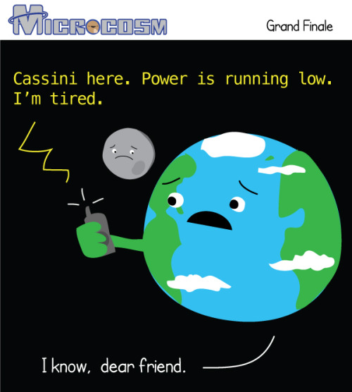
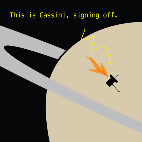
Friday, Cassini will dive into Saturn’s atmosphere and put an end to its nearly 20 year mission. Over those years we learned an incredible amount of information about Saturn, its rings, and its many moons. During the grand finale, Cassini will continue to send back information about Saturns atmosphere before burning up like a shooting star.

From vision to hand action
Our hands are highly developed grasping organs that are in continuous use. Long before we stir our first cup of coffee in the morning, our hands have executed a multitude of grasps. Directing a pen between our thumb and index finger over a piece of paper with absolute precision appears as easy as catching a ball or operating a doorknob. The neuroscientists Stefan Schaffelhofer and Hansjörg Scherberger of the German Primate Center (DPZ) have studied how the brain controls the different grasping movements. In their research with rhesus macaques, it was found that the three brain areas AIP, F5 and M1 that are responsible for planning and executing hand movements, perform different tasks within their neural network. The AIP area is mainly responsible for processing visual features of objects, such as their size and shape. This optical information is translated into motor commands in the F5 area. The M1 area is ultimately responsible for turning this motor commands into actions. The results of the study contribute to the development of neuroprosthetics that should help paralyzed patients to regain their hand functions (eLife, 2016).
The three brain areas AIP, F5 and M1 lay in the cerebral cortex and form a neural network responsible for translating visual properties of an object into a corresponding hand movement. Until now, the details of how this “visuomotor transformation” are performed have been unclear. During the course of his PhD thesis at the German Primate Center, neuroscientist Stefan Schaffelhofer intensively studied the neural mechanisms that control grasping movements. “We wanted to find out how and where visual information about grasped objects, for example their shape or size, and motor characteristics of the hand, like the strength and type of a grip, are processed in the different grasp-related areas of the brain”, says Schaffelhofer.
For this, two rhesus macaques were trained to repeatedly grasp 50 different objects. At the same time, the activity of hundreds of nerve cells was measured with so-called microelectrode arrays. In order to compare the applied grip types with the neural signals, the monkeys wore an electromagnetic data glove that recorded all the finger and hand movements. The experimental setup was designed to individually observe the phases of the visuomotor transformation in the brain, namely the processing of visual object properties, the motion planning and execution. For this, the scientists developed a delayed grasping task. In order for the monkey to see the object, it was briefly lit before the start of the grasping movement. The subsequent movement took place in the dark with a short delay. In this way, visual and motor signals of neurons could be examined separately.
The results show that the AIP area is primarily responsible for the processing of visual object features. “The neurons mainly respond to the three-dimensional shape of different objects”, says Stefan Schaffelhofer. “Due to the different activity of the neurons, we could precisely distinguish as to whether the monkeys had seen a sphere, cube or cylinder. Even abstract object shapes could be differentiated based on the observed cell activity.”
In contrast to AIP, area F5 and M1 did not represent object geometries, but the corresponding hand configurations used to grasp the objects. The information of F5 and M1 neurons indicated a strong resemblance to the hand movements recorded with the data glove. “In our study we were able to show where and how visual properties of objects are converted into corresponding movement commands”, says Stefan Schaffelhofer. “In this process, the F5 area plays a central role in visuomotor transformation. Its neurons receive direct visual object information from AIP and can translate the signals into motor plans that are then executed in M1. Thus, area F5 has contact to both, the visual and motor part of the brain.”
Knowledge of how to control grasp movements is essential for the development of neuronal hand prosthetics. “In paraplegic patients, the connection between the brain and limbs is no longer functional. Neural interfaces can replace this functionality”, says Hansjörg Scherberger, head of the Neurobiology Laboratory at the DPZ. “They can read the motor signals in the brain and use them for prosthetic control. In order to program these interfaces properly, it is crucial to know how and where our brain controls the grasping movements”. The findings of this study will facilitate to new neuroprosthetic applications that can selectively process the areas’ individual information in order to improve their usability and accuracy.
Newly discovered windows of brain plasticity may help with treatment of stress-related disorders
Chronic stress can lead to changes in neural circuitry that leave the brain trapped in states of anxiety and depression. But even under repeated stress, brief opportunities for recovery can open up, according to new research at The Rockefeller University.

(Image caption: Routine versus disruptive: A familiar stressor (left) did not increase NMDA receptors (dark spots), a booster of potentially harmful glutamate signaling, in the brains of mice. However, when subjected to an unfamiliar stress (right), mice expressed more NMDA receptors)
“Even after a long period of chronic stress, the brain retains the ability to change and adapt. In experiments with mice, we discovered the mechanism that alters expression of key glutamate-controlling genes to make windows of stress-related neuroplasticity—and potential recovery—possible,” says senior author Bruce McEwen, Alfred E. Mirsky Professor, and head of the Harold and Margaret Milliken Hatch Laboratory of Neuroendocrinology. Glutamate is a chemical signal implicated in stress-related disorders, including depression.
“This sensitive window could provide an opportunity for treatment, when the brain is most responsive to efforts to restore neural circuitry in the affected areas,” he adds.
The team, including McEwen and first author Carla Nasca, wanted to know how a history of stress could alter the brain’s response to further stress. To find out, they accustomed mice to a daily experience they dislike, confinement in a small space for a short period. On the 22nd day, they introduced some of those mice to a new stressor; others received the now-familiar confinement.
Then, the researchers tested both groups for anxiety- or depression-like behaviors. A telling split emerged: Mice tested shortly after the receiving the familiar stressor showed fewer of those behaviors; meanwhile those given the unfamiliar stressor, displayed more. The difference was transitory, however; by 24 hours after the final stressor, the behavioral improvements seen in half of the mice had disappeared.
Molecular analyses revealed a parallel fluctuation in a part of the hippocampus, a brain region involved in the stress response. A key molecule, mGlu2, which tamps down the release of the neurotransmitter glutamate, increased temporarily in mice subjected to the familiar confinement stress. Meanwhile, a molecular glutamate booster, NMDA, increased in other mice that experienced the unfamiliar stressor. In stress-related disorders, excessive glutamate causes harmful structural changes in the brain.
The researchers also identified the molecule regulating the regulator, an enzyme called P300. By adding chemical groups to proteins known as histones, which give support and structure to DNA, P300 increases expression of mGlu2, they found.
In other experiments, they looked at mice genetically engineered to carry a genetic variant associated with development of depression and other stress-related disorders in humans, and present in 33 percent of the population.
“Here again, in experiments relevant to humans, we saw the same window of plasticity, with the same up-then-down fluctuations in mGlu2 and P300 in the hippocampus,” Nasca says. “This result suggests we can take advantage of these windows of plasticity through treatments, including the next generation of drugs, such as acetyl carnitine, that target mGlu2—not to ‘roll back the clock’ but rather to change the trajectory of such brain plasticity toward more positive directions.”


Women scientists made up 25% of the Pluto fly-by New Horizon team. Make sure you share this, because erasing women’s achievements in science and history is a tradition. Happens every day.
.
http://pluto.jhuapl.edu/News-Center/News-Article.php?page=20150712

Scientists have discovered the world’s oldest known water in an ancient pool in Canada that’s at least 2 billion years old.
Back in 2013 they found water dating back about 1.5 billion years at the Kidd Mine in Ontario, but searching deeper at the site revealed an even older source buried underground.
The initial discovery of the ancient liquid in 2013 came at a depth of around 2.4 kilometres (1.5 miles) in an underground tunnel in the mine. But the extreme depth of the mine – which at 3.1 kilometres (1.9 miles) is the deepest base metal mine in the world – gave researchers the opportunity to keep digging.
“[The 2013 find] really pushed back our understanding of how old flowing water could be and so it really drove us to explore further,” geochemist Barbara Sherwood Lollar from the University of Toronto told Rebecca Morelle at the BBC.
“And we took advantage of the fact that the mine is continuing to explore deeper and deeper into the earth.”
The new source was found at about 3 kilometres (1.9 miles) down, and according to Sherwood Lollar, there’s a lot more of it than you might expect.
Continue Reading.
-
 science-is-magical reblogged this · 8 years ago
 -
 glitter-covered-thoughts reblogged this · 9 years ago
 -
 supermeganerd2012 liked this · 9 years ago
 -
 thebluecloudsflyup reblogged this · 9 years ago
 -
 thebluecloudsflyup liked this · 9 years ago
 -
 ghouldilocks liked this · 9 years ago
 -
 astro0livia reblogged this · 9 years ago
 -
 amihappytooihaventchecked liked this · 9 years ago
 -
 silverstargate liked this · 9 years ago
 -
 1998ish liked this · 9 years ago
 -
 insectoid5 liked this · 9 years ago
 -
 morgaine2005 reblogged this · 9 years ago
 -
 gofoxyourselves liked this · 9 years ago
 -
 cursedquill liked this · 9 years ago
 -
 ninjedithlord27 reblogged this · 9 years ago
 -
 chocaholicsanonymous liked this · 9 years ago
 -
 nothanksimgoodluv liked this · 9 years ago
 -
 periodicmemes liked this · 9 years ago
 -
 makitoujou reblogged this · 9 years ago
 -
 makitoujou liked this · 9 years ago
 -
 amdhaz liked this · 9 years ago
 -
 zakx2 reblogged this · 9 years ago
 -
 captainofthenx02 liked this · 9 years ago
 -
 turquoiseorchid reblogged this · 9 years ago
 -
 fanaa20 liked this · 9 years ago
 -
 sinkingmothership reblogged this · 9 years ago
 -
 newtonthinksimattractive reblogged this · 9 years ago
 -
 ddooyoung liked this · 9 years ago
 -
 that-relatable-demon-blog reblogged this · 9 years ago
 -
 that-relatable-demon-blog liked this · 9 years ago
 -
 vikuruh liked this · 9 years ago
 -
 teraleon liked this · 9 years ago
 -
 e-n-t-p liked this · 9 years ago
 -
 dinochickennugget reblogged this · 9 years ago
 -
 stonehengelesbian reblogged this · 9 years ago
 -
 tau-phi liked this · 9 years ago
 -
 cod-black reblogged this · 9 years ago
 -
 delinquentrawkkitty liked this · 9 years ago


