279 posts
Latest Posts by science-is-magical - Page 7

One of the best ways to get people to do something is to make it fun. A marketing campaign called ‘The Fun Theory’ found that when stairs next to an escalator were turned into giant, functional piano keys, the amount of people who took the stairs increased 66%, and when a trash can had sound effects that made it seem infinitely deep, people started picking up and throwing away litter just to hear it. Source


Drone with grabbing claw arms can lift 44 pounds
Prodrone’s latest creation could lift a four-year-old child, and uses its 5-axis metal claws to perch on fences like a bird.
Good news today: the fastest horizontal flyer in the animal kingdom is now a bat.
Free-tailed bats have now been clocked flying horizontally at over 160 kilometers per hour (that’s nearly 100 mph!), toppling the previous record-holder, the swift. The record for speed of diving is still held by the peregrine falcon but we’re coming for you next, feathers.
Source
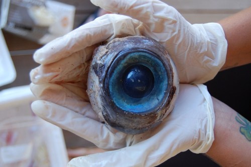
A giant eyeball from a mysterious sea creature was found by a man walking the beach in Pompano Beach on Wednesday. Wildlife officials said it likely came from a swordfish but experts mused that it likely came from a giant squid, whale or large fish. (Source)


How the Geneva Drive (the mechanical step that makes the second hand on a clock work by turning constant rotation into intermittent motion) works.

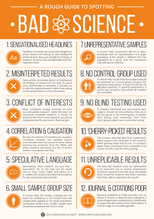
Version 1 of ‘A Rough Guide to Spotting Bad Science’. Thanks for everyone’s suggestions earlier in the week, attempted to include as many of them as possible!
Download link here: http://wp.me/p4aPLT-ap

Scientists use lasers to control mouse brain switchboard
Ever wonder why it’s hard to focus after a bad night’s sleep? Using mice and flashes of light, scientists show that just a few nerve cells in the brain may control the switch between internal thoughts and external distractions. The study, partly funded by the National Institutes of Health, may be a breakthrough in understanding how a critical part of the brain, called the thalamic reticular nucleus (TRN), influences consciousness.
“Now we may have a handle on how this tiny part of the brain exerts tremendous control over our thoughts and perceptions,” said Michael Halassa, M.D., Ph.D., assistant professor at New York University’s Langone Medical Center and a lead investigator of the study. “These results may be a gateway into understanding the circuitry that underlies neuropsychiatric disorders.”
The TRN is a thin layer of nerve cells on the surface of the thalamus, a center located deep inside the brain that relays information from the body to the cerebral cortex. The cortex is the outer, multi-folded layer of the brain that controls numerous functions, including one’s thoughts, movements, language, emotions, memories, and visual perceptions. TRN cells are thought to act as switchboard operators that control the flow of information relayed from the thalamus to the cortex.
“The future of brain research is in studying circuits that are critical for brain health and these results may take us a step further,” said James Gnadt, Ph.D., program director at NIH’s National Institute Neurological Disorders and Stroke (NINDS), which helped fund the study. “Understanding brain circuits at the level of detail attained in this study is a goal of the President’s Brain Research through Advancing Innovative Neurotechnologies (BRAIN) Initiative.”
To study the circuits, the researchers identified TRN cells that send inhibitory signals to parts of the thalamus known to relay visual information to the cortex. Using a technique known as multi-electrode recordings, they showed that sleep and concentration affected these cells in opposite ways.
They fired often when the mice were asleep, especially during short bursts of simultaneous brain cell activity called sleep spindles. These activity bursts briefly widen electrical brain wave traces making them look like spindles, the straight spikes with rounded bottoms used to make yarn. In contrast, the cells fired infrequently when the mice were tasked with using visual cues to find food. The results suggested that these cells blocked visual information from reaching the cortex during sleep and allowed its transmission when the mice were awake and attentive.
For Dr. Halassa, a practicing psychiatrist who treats schizophrenia, these surprising results may provide fundamental insights into how the brain controls information transmission, a process that is disrupted in patients with neuropsychiatric disorders. Previous studies suggested that people who experienced more spindles while sleeping were less susceptible to being disturbed by outside noises. Moreover, people with schizophrenia and autism spectrum disorder may experience fewer spindles.
“Spindles may be peepholes into the mysteries of these disorders,” said Dr. Halassa.
To test this idea, the researchers used optogenetics, a technique that introduces light-sensitive molecules into nerve cells. This allowed them to precisely control the firing patterns of visual TRN cells with flashes of laser light. The experiments were performed in well-rested as well as sleep-deprived mice. Similar to what is seen in humans, sleep deprivation can disrupt the ability of mice to focus and block out external distractions.
Well-rested mice needed just a second or two to find the food whereas sleep-deprived mice took longer, suggesting that lack of sleep had detrimental effects on their ability to focus. When the researchers used flashes of laser light to inhibit the firing of optogenetically engineered visual TRN cells in sleep-deprived mice, the mice found the food faster. In contrast, if they used optogenetics to induce sleep-like firing patterns in well-rested mice, then the mice took longer to find food.
“It’s as if with a flick of a switch we could alter the mental states of the mice and either mimic or cure their drowsiness,” said Dr. Halassa.
In a parallel set of experiments the researchers found neighbors of the visual TRN cells had very different characteristics. These neighboring cells control the flow of information to the cortex from limbic brain regions, which are involved with memory formation, emotions and arousal. The cells fired very little during sleep and instead were active when the mice were awake. Dr. Halassa thinks that their firing pattern may be important for the strengthening of new memories that often occurs during sleep. Combined, the results suggest that the TRN is divided into sub-networks that oversee discrete mental states. The researchers think understanding the sub-networks is an initial step in thoroughly exploring the role of the TRN in brain disorders.
Ok, so I don’t know how I ended up here and woah!
they made

characters

for

every

single

element

of the

periodic

table!

And also they made this

and this

*new ship*
There’s even a granny!

It’s like

superheros

(there’s a guy who looks like Hulk btw)

and humans

and there are

twins!!

And Bethoveen

THEY MADE THOR

And there’s also this which made me laugh

I can’t!

(source)
This is the chemical formula for love: C8H11NO2+C10H12N2O+C43H66N12O12S2 dopamine, seratonin, oxytocin. It can be easily manufactured in a lab, but overdosing on any of them can cause schizophrenia, extreme paranoia, and insanity. Let that sink in.
(via mrzim)


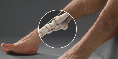
Redrawing the brain’s motor map
Neuroscientists at Emory have refined a map showing which parts of the brain are activated during head rotation, resolving a decades-old puzzle. Their findings may help in the study of movement disorders affecting the head and neck, such as cervical dystonia and head tremor.
The results were published in Journal of Neuroscience.
In landmark experiments published in the 1940s and 50s, Canadian neurosurgeon Wilder Penfield and colleagues determined which parts of the motor cortex controlled the movements of which parts of the body.
Penfield stimulated the brain with electricity in patients undergoing epilepsy surgery, and used the results to draw a “motor homunculus”: a distorted representation of the human body within the brain. Penfield assigned control of the neck muscles to a region between those that control the fingers and face, a finding inconsistent with some studies that came later.
Using modern functional MRI (magnetic resonance imaging), researchers at Emory University School of Medicine have shown that the neck’s motor control region in the brain is actually between the shoulders and trunk, a location that more closely matches the arrangement of the body itself.
“We can’t be that hard on Penfield, because the number of cases where he was able to study head movement was quite limited, and studying head motion as he did, by applying an electrode directly to the brain, creates some challenges,” says lead author Buz Jinnah, MD, professor of neurology, human genetics and pediatrics at Emory University School of Medicine.
The new location for the neck muscles makes more sense, because it corresponds to a similar map Penfield established of the sense of touch (the somatosensory cortex), Jinnah says.
Participants in brain imaging studies need to keep their heads still to provide accurate data, so volunteers were asked to perform isometric muscle contraction. They attempted to rotate their heads to the left or the right, even though head movement was restricted by foam padding and restraining straps.
First author Cecilia Prudente, a graduate student in neuroscience who is now a postdoctoral associate at the University of Minnesota, developed the isometric head movement task and obtained internal funding that allowed the study to proceed.
She and Jinnah knew that isometric exercises for the wrist activated the same regions of the motor cortex as wrist movements, and used that as a reference point in their study. During brain imaging, they were able to check that particular muscles were being tensed by directly monitoring volunteers’ muscles electronically.
When volunteers contracted their neck muscles, researchers were able to detect activation in other parts of the brain too, such as the cerebellum and the basal ganglia, which are known to be involved in movement control. This comes as no surprise, Jinnah says, since these regions also control movements of the hands and other body parts.
Prudente, Jinnah and colleagues have conducted a similar study with cervical dystonia patients, with the goal of comparing the patterns of brain activation between healthy volunteers and the patients. Cervical dystonia is a painful condition in which the neck muscles contract involuntarily and the head posture is distorted.
“These results may help guide future studies in humans and animals, as well as medical or surgical interventions for cervical dystonia and other disorders involving abnormal head movements,” Prudente says.


Photographs taken of Saturn by NASA. Yes, these are real pictures; they are not illustrations.
(Source)
A puzzling expected value
Pick a (uniformly) random real number from the unit interval [0,1] and repeat this until the sum of all chosen numbers exceeds 1. What is the expected number of real values you will pick?
The quite surprising answer is Eulers constant, e ≈ 2.71828.
A demonstration can be found on Wolfram MathWorld.


Here’s how nightmarish humans would look if our bodies were designed to survive car crashes
Article by Chris Weller, Tech Insider & Business Insider
If you’re ever in a car with Graham, then don’t bother telling him to buckle his seat belt. His body is already designed to withstand high-speed impacts.
Designed by a trauma surgeon, an artist, and a crash investigator, Graham is a hypothetical scenario come to life. Supported by Australia’s Transport Accident Commission, the project is meant to highlight how vulnerable humans are to injury.
Graham, however, is not.
Keep reading

About 400 million years ago, before trees were common, the Earth was covered with giant mushrooms. Source
Neuroscientist Discovers Potential New Source for Pain Inhibition
A UT Dallas scientist has found a new neurological mechanism that appears to contribute to a reduction in pain.
According to Dr. Ted Price, associate professor in the School of Behavioral and Brain Sciences, the discovery of neuroligin-2 as a cause exacerbating chronic pain is significant for the research community. Although the findings likely won’t immediately lead to new pain therapies, the findings offer a potential new therapeutic direction to investigate, he said.
Price’s research on the topic has recently been published online in Pain, the journal of the International Association for the Study of Pain.
The study focused on the body’s inhibitory networks — a series of biochemical reactions that decrease certain neurological activity, such as pain. Price said a great deal of previous research in this area has focused on the activity of the neurotransmitter GABA, a chemical released by nerve cells in the brain.
Normally, a GABA neurotransmitter acts to inhibit neuronal activity, such as pain. However, when pain becomes chronic there is strong evidence that a process called GABAergic plasticity can cause GABA to lose its inhibitory activity, sometimes making the pain even worse.
The source of these excitatory actions in neuronal circuits has been broadly attributed to chloride ions, but Price’s research has found another potential cause of GABAergic plasticity: synaptic adhesion molecules called neuroligin-2.
“From a basic science perspective, we’re really excited about it because it demonstrates that the types of GABAergic plasticity that can occur in the setting of chronic pain are more diverse than we’ve appreciated before,” he said.
Price, who heads the undergraduate research program in neuroscience in the school, focuses much of his research on understanding the neuroscience behind pain, particularly chronic pain. He said individuals with chronic pain typically don’t receive the pain-reduction benefits delivered by inhibitory systems. Instead, they often experience increased pain.
“When you hit your hand with a hammer, almost everybody has the same reflex reaction — that is, to rub your finger which, in turn, helps to reduce pain. The reason that works is because it increases GABAergic inhibition in the spinal cord,” Price said. “However, people who have chronic pain — if they do the same thing — find that rubbing it actually makes the pain worse. That’s because the GABAergic system loses its efficacy and, in fact, can become excitatory.”
Price said the research is another step in determining why the GABAergic system stops working correctly in some people and provides a second theory for what drives the system.
“Having two ideas and different models will allow us to determine what the therapeutic opportunities are — creating something that will change that back to normal. The lack of performance in the inhibitory system is very detrimental to those who are in chronic pain,” he said.
Price said the development of chronic pain is, in essence, one’s body “learning” something that is bad.
“It’s changing the way the body functions — it’s learning. That learning, in the case of chronic pain, is aberrant — it’s causing the situation to get worse. If we can figure out what that form of learning was, then we can potentially reverse it. Understanding that the GABAergic system changes during this form of learning potentially offers a new therapeutic avenue,” he said.


How Printing a 3-D Skull Helped Save a Real One
What started as a stuffy-nose and mild cold symptoms for 15-year-old Parker Turchan led to a far more serious diagnosis: a rare type of tumor in his nose and sinuses that extended through his skull near his brain.
“He had always been a healthy kid, so we never imagined he had a tumor,” says Parker’s father, Karl. “We didn’t even know you could get a tumor in the back of your nose.”
The Portage, Michigan, high school sophomore was referred to the University of Michigan’s C.S. Mott Children’s Hospital, where doctors determined the tumor extended so deep that it was beyond what regular endoscopy could see.
The team members needed to get the best representation of the tumor’s extent to ensure that their surgical approach could successfully remove the entire mass
“Parker had an uncommon, large, high-stage tumor in a very challenging area,” says Mott pediatric head and neck surgeon David Zopf, M.D. “The tumor’s location and size had me question whether a minimally invasive approach would allow us to remove the tumor completely.”
To help answer that question, teams at Mott sought an innovative approach: crafting a 3-D replica of Parker’s skull.
The model, made of polylactic acid, helped simulate the coming operation on Parker by giving U-M surgeons “an exact replica of his craniofacial anatomy and a way to essentially touch the ‘tumor’ with our hands ahead of time,” Zopf says.
Just as important, it also allowed the team to counsel Parker and his family by offering them a look at what lurked within — and, with the test run successfully complete, what would lie ahead.
A ‘pretty impressive’ model
The rare and aggressive tumor in Parker’s nose is known as juvenile nasopharyngeal angiofibroma, a mass that grows in the back of the nasal cavity and predominantly affects young male teens. Mott sees a handful of cases each year.
In Parker’s case, the tumor had two large parts: one roughly the size of an egg and the other the size of a kiwi. The mass sat right in the center of the craniofacial skeleton below the brain and next to the nerves that control eye movement and vision.
“We were obviously concerned about the risks involved in this kind of procedure, which we knew could lead to a lot of blood loss and was sensitive because it was so close to the nerves in his face,” says Karl, who praised the 3-D methodology used to aid his son. “It was pretty impressive to see the model of Parker’s skull ahead of the surgery. We had no idea this was even possible.”
Zopf, working with Erin McKean, M.D., a U-M skull base surgeon, was able to completely remove the large tumor. Kyle VanKoevering, M.D., and Sajad Arabnejad, Ph.D., aided in model preparation.
Through preoperative embolization, the blood supply to the tumor was blocked off the day before surgery to decrease blood loss. A large portion of the tumor was then detached endoscopically and removed through the mouth. The remaining mass under the brain was taken out through the nose.
Doctors took pictures of Parker’s anatomy during the surgery and, later, compared it with pictures from the model. They were nearly identical.
“Words alone can’t express how thankful we are for Parker’s talented team of surgeons at Mott,” says his mother, Heidi. “Parker is back to his old self again.”
Powerful potential
Although medical application of the technology continues to gain attention, it isn’t entirely new. Zopf and Mott teams have used 3-D printing for almost five years.
Groundbreaking 3-D printed splints made at U-M have helped save the lives of babies with severe tracheobronchomalacia, which causes the windpipe to periodically collapse and prevents normal breathing. Mott has also used 3-D printing on a fetus to plan for a potentially complicated birth.
“We are finding more and more uses for 3-D printing in medicine,” Zopf says. “It is proving to be a powerful tool that will allow for enhanced patient care.”
Based on success in patients such as Parker and continued collaboration, it’s a concept that appears poised to thrive.
“Because of the team approach we’ve established at the University of Michigan between otolaryngology and biomedical engineering, the printed models can be designed and rapidly produced at a very low cost,” Zopf says. “Michigan is one of only a few places in the nation and world that has the capacity to do this.”
Your body is an incredibly bizarre machine.
“What you see is a myosin protein dragging an endorphin along a filament to the inner part of the brain’s parietal cortex which creates happiness. Happiness. You’re looking at happiness.”

My tummy is blushing now.
People Were Asked: ‘What’s The Coolest Thing Most People Don’t Know About Their Own Body?’
Listening To Music Releases Dopamine In The Brain
Have you ever been listening to a piece of music and experienced intense pleasure, even chills? Valorie Salimpoor and team (2010) conducted research that shows that listening to music can release the neurotransmitter dopamine.
A wide range of music — The researchers used PET (positron emission tomography) scans, fMRI, and psychophysiological measures such as heart rate to measure reactions while people listened to music. The participants provided music that they said gave them intense pleasure and chills. The range of music varied, from classical, folk, jazz, elecronica, rock pop, tango, and more.
Keep reading