Tag Later - Blog Posts


Happy TRAPPIST-1 Day!
Here’s a comic on our latest discovery!
http://www.space.com/35806-trappist-1-facts.html

Total breakdown in the brain
A stroke is just one example of a condition when communication between nerve cells breaks down. Micro-failures in brain functioning also occur in conditions such as depression and dementia. In most cases, the lost capacity will return after a while. However, consequential damage will often remain so that the functional capability can only be restored through lengthy treatment — if at all. For this reason, researchers at Friedrich-Alexander-Universität Erlangen-Nürnberg (FAU) have been investigating what happens during such breakdown phases and looking at possible ways of preventing damage and speeding up the healing processes. Their findings have been recently published in the eminent journal Scientific Reports.

(Image caption: Nerve cell networks visualised using high-speed fluorescent microscopy and then reconstructed with the software developed by Wrosch and her team. Credit: FAU/Jana Wrosch)
The research team headed by Jana Wrosch of FAU’s Chair of Psychiatry and Psychotherapy found that significant alterations occurred in neural cells while the communication pathways were blocked. Neuron networks reconnect during such periods of inactivity and become hypersensitive. If we imagine that normal communication pathways are motorways, when they are blocked a form of traffic chaos occurs in the brain whereby information is re-routed in disorganised form along what can be called side streets and minor routes. Additional synapses are generated everywhere and begin operating. When the signal is reinstated, the previously coordinated information routes no longer exist and, as in the case of a child, the appropriate functions need to be learned from scratch. Since they are receiving no normal signals during the phase of brain malfunction, the nerve cells also become more sensitive in an attempt to find the missing input. Once the signals return, this means they may overreact.
Nerve cells flicker when stained
Visualising the microscopically minute connections between the nerve cells is a major technical challenge. The conventional microscopic techniques currently available, such as electron microscopy, always require preliminary treatment of the nerve cells that are to undergo examination. However, this causes the nerve cells to die, so that the alterations that occur in the cells cannot be observed. To get round this problem, Wrosch and her team have developed a high-speed microscopy process along with special statistical computer software that make it possible to visualise the communication networks of living neurons. First, a video of the cells is made whereby an image is taken every 36 milliseconds. A special dye is used to stain the cells to ensure that the individual cells flicker whenever they receive a signal. Subsequently, the software recognises these cells on the video images and detects the information pathways by which the signals are transmitted from cell to cell.
The nerve cells are then exposed to the pufferfish poison tetrodotoxin to simulate the blocking of communication channels that occurs in disorders. After inducing communication breakdown phases of varying lengths, the researchers remove the toxin from the cells and determine how the nerve cell networks have changed during exposure. ‘Thanks to this concept, we have been finally able to discover what happens when communication is blocked,’ explains Wrosch. ‘Now we can try to develop medications that will help prevent these damaging changes.’ In future projects, the research team plans to examine the exact mode of action of anti-depressants on nerve cell networks and intends to find new approaches to creating more effective drugs.

(Image caption: The above image compares the neural activation patterns between images from the participants’ brains when reading “O eleitor foi ao protesto” (observed) and the computational model’s prediction for “The voter went to the protest” (predicted))
Brain “Reads” Sentences the Same in English and Portuguese
An international research team led by Carnegie Mellon University has found that when the brain “reads” or decodes a sentence in English or Portuguese, its neural activation patterns are the same.
Published in NeuroImage, the study is the first to show that different languages have similar neural signatures for describing events and scenes. By using a machine-learning algorithm, the research team was able to understand the relationship between sentence meaning and brain activation patterns in English and then recognize sentence meaning based on activation patterns in Portuguese. The findings can be used to improve machine translation, brain decoding across languages and, potentially, second language instruction.
“This tells us that, for the most part, the language we happen to learn to speak does not change the organization of the brain,” said Marcel Just, the D.O. Hebb University Professor of Psychology and pioneer in using brain imaging and machine-learning techniques to identify how the brain deciphers thoughts and concepts.
“Semantic information is represented in the same place in the brain and the same pattern of intensities for everyone. Knowing this means that brain to brain or brain to computer interfaces can probably be the same for speakers of all languages,” Just said.
For the study, 15 native Portuguese speakers — eight were bilingual in Portuguese and English — read 60 sentences in Portuguese while in a functional magnetic resonance imaging (fMRI) scanner. A CMU-developed computational model was able to predict which sentences the participants were reading in Portuguese, based only on activation patterns.
The computational model uses a set of 42 concept-level semantic features and six markers of the concepts’ roles in the sentence, such as agent or action, to identify brain activation patterns in English.
With 67 percent accuracy, the model predicted which sentences were read in Portuguese. The resulting brain images showed that the activation patterns for the 60 sentences were in the same brain locations and at similar intensity levels for both English and Portuguese sentences.
Additionally, the results revealed the activation patterns could be grouped into four semantic categories, depending on the sentence’s focus: people, places, actions and feelings. The groupings were very similar across languages, reinforcing the organization of information in the brain is the same regardless of the language in which it is expressed.
“The cross-language prediction model captured the conceptual gist of the described event or state in the sentences, rather than depending on particular language idiosyncrasies. It demonstrated a meta-language prediction capability from neural signals across people, languages and bilingual status,” said Ying Yang, a postdoctoral associate in psychology at CMU and first author of the study.

Cassini has finally reached its end. But the fiery end doesn’t have to be as sad as we’ve imagined it. 🚀♥️ ✏️: @erika.nesvold #science #saturn #cassini #sad #home #sciencealert #beautiful http://ift.tt/2y7bUO1

Physicists Detect Gravitational Waves, Proving Einstein Right
On Thursday (Feb. 11, 2016) at 10:30 a.m. ET, the National Science Foundation will gather scientists from Caltech, MIT and the LIGO Scientific Collaboration in Washington D.C. to update the scientific community on the efforts being made by the Laser Interferometer Gravitational-wave Observatory (LIGO) to detect gravitational waves.
But why is this exciting? And what the heck are “gravitational waves”?
Keep reading


Bonus comic!
Yahoo! Einstein was right again! :D We now have our first detection of gravitational waves!
http://www.nytimes.com/2016/02/12/science/ligo-gravitational-waves-black-holes-einstein.html?_r=0
http://www.space.com/17661-theory-general-relativity.html


Newborn baby brain scans will help scientists track brain development
Scientists have published ground-breaking scans of newborn babies’ brains which researchers from all over the world can download and use to study how the human brain develops.

(Image caption: Diffusion MRI of the developing neonatal brain (Left: Multi-shell high angular resolution diffusion data decomposed into a free water component (greyscale background image) and a directionally resolved brain tissue component shown as rendered surfaces. Middle and right: Visualisation of anatomical connections in the developing brain derived from the brain tissue component.)
The images are part of the Developing Human Connectome Project (dHCP), a collaboration between King’s College London, Imperial College London and the University of Oxford, which will uncover how the brain develops, including the wiring and function of the brain during pregnancy and how this changes after birth. The dHCP researchers are sharing their images and methods online so that other scientists from around the world can use the data in their own research.
Using Magnetic Resonance Imaging (MRI) scanners at Evelina London Children’s Hospital, the team has developed new techniques which enable images of the brains of foetuses and babies to be captured. Researchers have overcome problems caused by babies’ movement and small size, as well as the difficulties in keeping vulnerable infants safe in the MRI scanner, so that they can now produce highly detailed and rich information on brain development.
The project will help scientists to understand how conditions such as autism develop, or how problems in pregnancy affect brain growth.
‘The Developing Human Connectome Project is a major advance in understanding human brain development - it will provide the first map of how the brain’s connections develop, and how this goes wrong in disease,’ said Lead Principal Investigator, Professor David Edwards from King’s College London and Consultant Neonatologist at Evelina London.
The research collaboration is funded by a €15 million Synergy Grant from the European Research Council, and one of the goals of the project is to make sure that the data is shared as widely across the world as possible. Scientists are able to download the images at https://data.developingconnectome.org.
For this project, Professor Jo Hajnal’s team at King’s College London developed new MRI technology specifically designed to provide high resolution scans of newborn and fetal brains.
In addition, a group led by Professor Daniel Rueckert’s at Imperial College London developed new computer programs to analyse the images. 'We have been developing novel approaches that help researchers by automatically analysing the rich and comprehensive MR images that are collected as part of dHCP,’ he explained.
At the University of Oxford, Professor Steve Smith’s team has been developing specific techniques to define where the connections are in the developing brain.
As well as studying more babies, the team at Evelina London are now recruiting pregnant mothers for foetal scanning. Meanwhile, the first release of images today will allow scientists to start to explore these powerful images and begin mapping out the complexities of human brain development in a whole new way.
Magnetospheres: How Do They Work?
The sun, Earth, and many other planets are surrounded by giant magnetic bubbles.

Space may seem empty, but it’s actually a dynamic place, dominated by invisible forces, including those created by magnetic fields. Magnetospheres – the areas around planets and stars dominated by their magnetic fields – are found throughout our solar system. They deflect high-energy, charged particles called cosmic rays that are mostly spewed out by the sun, but can also come from interstellar space. Along with atmospheres, they help protect the planets’ surfaces from this harmful radiation.
It’s possible that Earth’s protective magnetosphere was essential for the development of conditions friendly to life, so finding magnetospheres around other planets is a big step toward determining if they could support life.
But not all magnetospheres are created equal – even in our own backyard, not all planets in our solar system have a magnetic field, and the ones we have observed are all surprisingly different.

Earth’s magnetosphere is created by the constantly moving molten metal inside Earth. This invisible “force field” around our planet has an ice cream cone-like shape, with a rounded front and a long, trailing tail that faces away from the sun. The magnetosphere is shaped that way because of the constant pressure from the solar wind and magnetic fields on the sun-facing side.

Earth’s magnetosphere deflects most charged particles away from our planet – but some do become trapped in the magnetic field and create auroras when they rain down into the atmosphere.

We have several missions that study Earth’s magnetosphere – including the Magnetospheric Multiscale mission, Van Allen Probes, and Time History of Events and Macroscale Interactions during Substorms (also known as THEMIS) – along with a host of other satellites that study other aspects of the sun-Earth connection.


Mercury, with a substantial iron-rich core, has a magnetic field that is only about 1% as strong as Earth’s. It is thought that the planet’s magnetosphere is stifled by the intense solar wind, limiting its strength, although even without this effect, it still would not be as strong as Earth’s. The MESSENGER satellite orbited Mercury from 2011 to 2015, helping us understand our tiny terrestrial neighbor.


After the sun, Jupiter has by far the biggest magnetosphere in our solar system – it stretches about 12 million miles from east to west, almost 15 times the width of the sun. (Earth’s, on the other hand, could easily fit inside the sun.) Jupiter does not have a molten metal core like Earth; instead, its magnetic field is created by a core of compressed liquid metallic hydrogen.

One of Jupiter’s moons, Io, has intense volcanic activity that spews particles into Jupiter’s magnetosphere. These particles create intense radiation belts and the large auroras around Jupiter’s poles.

Ganymede, Jupiter’s largest moon, also has its own magnetic field and magnetosphere – making it the only moon with one. Its weak field, nestled in Jupiter’s enormous shell, scarcely ruffles the planet’s magnetic field.
Our Juno mission orbits inside the Jovian magnetosphere sending back observations so we can better understand this region. Previous observations have been received from Pioneers 10 and 11, Voyagers 1 and 2, Ulysses, Galileo and Cassini in their flybys and orbits around Jupiter.

Saturn’s moon Enceladus transforms the shape of its magnetosphere. Active geysers on the moon’s south pole eject oxygen and water molecules into the space around the planet. These particles, much like Io’s volcanic emissions at Jupiter, generate the auroras around the planet’s poles. Our Cassini mission studies Saturn’s magnetic field and auroras, as well as its moon Enceladus.


Uranus’ magnetosphere wasn’t discovered until 1986 when data from Voyager 2’s flyby revealed weak, variable radio emissions. Uranus’ magnetic field and rotation axis are out of alignment by 59 degrees, unlike Earth’s, whose magnetic field and rotation axis differ by only 11 degrees. On top of that, the magnetic field axis does not go through the center of the planet, so the strength of the magnetic field varies dramatically across the surface. This misalignment also means that Uranus’ magnetotail – the part of the magnetosphere that trails away from the sun – is twisted into a long corkscrew.


Neptune’s magnetosphere is also tilted from its rotation axis, but only by 47. Just like on Uranus, Neptune’s magnetic field strength varies across the planet. This also means that auroras can be seen away from the planet’s poles – not just at high latitudes, like on Earth, Jupiter and Saturn.

Does Every Planet Have a Magnetosphere?
Neither Venus nor Mars have global magnetic fields, although the interaction of the solar wind with their atmospheres does produce what scientists call an “induced magnetosphere.” Around these planets, the atmosphere deflects the solar wind particles, causing the solar wind’s magnetic field to wrap around the planet in a shape similar to Earth’s magnetosphere.

What About Beyond Our Solar System?
Outside of our solar system, auroras, which indicate the presence of a magnetosphere, have been spotted on brown dwarfs – objects that are bigger than planets but smaller than stars.
There’s also evidence to suggest that some giant exoplanets have magnetospheres. As scientists now believe that Earth’s protective magnetosphere was essential for the development of conditions friendly to life, finding magnetospheres around exoplanets is a big step in finding habitable worlds.
Make sure to follow us on Tumblr for your regular dose of space: http://nasa.tumblr.com

Researchers at King’s College London found that the drug Tideglusib stimulates the stem cells contained in the pulp of teeth so that they generate new dentine – the mineralised material under the enamel.
Teeth already have the capability of regenerating dentine if the pulp inside the tooth becomes exposed through a trauma or infection, but can only naturally make a very thin layer, and not enough to fill the deep cavities caused by tooth decay.
But Tideglusib switches off an enzyme called GSK-3 which prevents dentine from carrying on forming.
Scientists showed it is possible to soak a small biodegradable sponge with the drug and insert it into a cavity, where it triggers the growth of dentine and repairs the damage within six weeks.
The tiny sponges are made out of collagen so they melt away over time, leaving only the repaired tooth.

The existence of large numbers of molecules in winds powered by supermassive black holes at the centers of galaxies has puzzled astronomers since they were discovered more than a decade ago. Molecules trace the coldest parts of space, and black holes are the most energetic phenomena in the universe, so finding molecules in black hole winds was like discovering ice in a furnace.
Astronomers questioned how anything could survive the heat of the energetic outflows, but a new theory from researchers in Northwestern University’s Center for Interdisciplinary Research and Exploration in Astrophysics (CIERA) predicts that these molecules are not survivors at all, but brand-new molecules, born in the winds with unique properties that enable them to adapt to and thrive in the hostile environment.
Continue Reading.
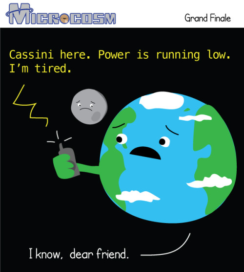
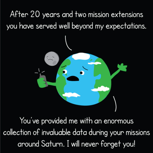


Friday, Cassini will dive into Saturn’s atmosphere and put an end to its nearly 20 year mission. Over those years we learned an incredible amount of information about Saturn, its rings, and its many moons. During the grand finale, Cassini will continue to send back information about Saturns atmosphere before burning up like a shooting star.
After 20 years in space, the Cassini spacecraft is running out of fuel. In 2010, Cassini began a seven-year mission extension in which the plan was to expend all of the spacecraft’s propellant exploring Saturn and its moons. This led to the Grand Finale and ends with a plunge into the planet’s atmosphere at 6:32 a.m. EDT on Friday, Sept. 15.
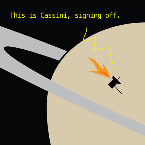
The spacecraft will ram through Saturn’s atmosphere at four times the speed of a re-entry vehicle entering Earth’s atmosphere, and Cassini has no heat shield. So temperatures around the spacecraft will increase by 30-to-100 times per minute, and every component of the spacecraft will disintegrate over the next couple of minutes…
Cassini’s gold-colored multi-layer insulation blankets will char and break apart, and then the spacecraft’s carbon fiber epoxy structures, such as the 11-foot (3-meter) wide high-gain antenna and the 30-foot (11-meter) long magnetometer boom, will weaken and break apart. Components mounted on the outside of the central body of the spacecraft will then break apart, followed by the leading face of the spacecraft itself.
Make sure to follow us on Tumblr for your regular dose of space: http://nasa.tumblr.com.


Words matter when talking about Alzheimer’s
Using war metaphors in reference to Alzheimer’s disease should be replaced with messages of resilience against a complex, age-associated condition that may not be fully defeatable, according to a team of researchers.
Framing a health issue through comparisons to warfare is common in popular media and medical and research communities. While it can motivate efforts to deal with the issue, this type of language and messaging can also create fear and stigma, turn patients into victims and divert resources from critically important prevention and care, said Daniel R. George, assistant professor of medical humanities, Penn State College of Medicine.
Despite decades of failures in Alzheimer’s drug development, scientific attention continues to focus on drugs that “attack” a molecular compound called beta amyloid, with the goal of curing the disease. Amyloid is a key component of the plaques in the brain that are a hallmark of Alzheimer’s disease. Research, however, shows that the appearance of amyloid does not correlate with clinical symptoms and beta amyloid has repeatedly been found in the brains of one-third of “normal” elderly people. This suggests that amyloid may be a symptom rather than a cause of damage. A growing number of researchers believe that declaring “war” on Alzheimer’s by “attacking” amyloid may ultimately be an exercise in self-harm, particularly if amyloid is representative of the brain’s repair response, and may be channeling resources away from other drug-based approaches that do not assume amyloid toxicity.
Scholars have argued that metaphors and narratives that treat disease as something to be attacked can be socially damaging to those affected. The value of such metaphors may be clearer for infectious diseases caused by single pathogens. It becomes more problematic when discussing diverse, age-associated syndromes like Alzheimer’s that may not be fully curable. In this way, war metaphors in medicine can invite ways of thinking that may not be scientifically or socially productive.
“If applied in a careless manner, war metaphors can delude our sense of what’s possible therapeutically, and give false hope to people and caregivers who are suffering,” George said.
George and his co-authors propose moving toward different types of metaphors – those that encourage use of words like “slow” or “postpone” rather than “prevent” or “cure,” and emphasize building “resilience” to aging processes in the brain rather than aiming at “absolute victory” over a disease. While “fighting” and “defeating” Alzheimer’s through drug development is important, the authors argue it may be wiser to acknowledge that Alzheimer’s is not a disease disconnected from the aging process like polio or malaria. The authors note that Alzheimer’s has been classified as a disease for the past 40 years. They suggest it may be more beneficial to take a lifespan-oriented approach that includes education about known biological, psychosocial and environmental risk factors, investment in societal programs and infrastructure that support brain health, and ensuring proper care for those affected and their caregivers.
“While not as profitable as drug development, public health initiatives that reduce vascular risk factors, modulate oxidative stress and inflammation, guard against traumatic brain injuries, promote social engagement and lifelong learning, and reduce exposure to neurotoxins, and other commonsense actions should be an explicit component of our societal response (to Alzheimer’s),” the researchers wrote in the American Journal of Bioethics.
George drew particular attention to the residents of Flint, Michigan being exposed to lead, a neurotoxin, through the water supply.
“It is inexcusable that we could let our public infrastructure fail to the point where it becomes a contributor to Alzheimer’s disease risk for socio-economically disadvantaged citizens,” George said. “If we’re really serious about addressing the problem of Alzheimer’s, we must start by not poisoning our citizens.”
Moving beyond the notion of being at war against Alzheimer’s could also serve to humanize cognitive aging.
“There’s a widely-accepted myth that people who have Alzheimer’s are sort of non-people, akin to zombies,” George said. “There are ways to construct meaning around memory loss that show greater compassion and solidarity toward people with cognitive frailty rather than seeing them as passive victims in our biological war against the disease. We believe in a more humane message – that even if you have a diagnosis of ‘probable Alzheimer’s’ you can still have a life with deep purpose, social contribution and meaningful relationships.”

I am not going to tag the name of the bird, because I’m pretty sure I would get tagged as NSFW if I did, but I assure you their beaks are getting longer and it’s probably because of the UK’s obsession with bird feeders.


After the Battle of Shiloh in 1862, many Civil War soldiers’ lives were saved by a phenomenon called ‘Angel’s Glow.’ The soldiers, who lay in the mud for two rainy days, had wounds that began to glow in the dark and heal unusually fast. In 2001, 2 teens won an international science fair by discovering the soldiers had been so cold that their bodies created the perfect conditions for growing a bioluminescent bacteria, which ultimately destroyed the bad bacteria that could’ve killed them. Source Source 2 Source 3


Francium (new video) - Periodic Table of Videos
Interesting video on the element Francium and its discoverer Marguerite Perey.
Flying to New Heights With the Magnetospheric Multiscale Mission
A mission studying Earth’s magnetic field by flying four identical spacecraft is headed into new territory.

The Magnetospheric Multiscale mission, or MMS, has been studying the magnetic field on the side of Earth facing the sun, the day side – but now we’re focusing on something else. On February 9, MMS started the three-month-long process of shifting to a new orbit.

One key thing MMS studies is magnetic reconnection – a process that occurs when magnetic fields collide and re-align explosively into new positions. The new orbit will allow MMS to study reconnection on the night side of the Earth, farther from the sun.

Magnetic reconnection on the night side of Earth is thought to be responsible for causing the northern and southern lights.

To study the interesting regions of Earth’s magnetic field on the night side, the four MMS spacecraft are being boosted into an orbit that takes them farther from Earth than ever before. Once it reaches its final orbit, MMS will shatter its previous Guinness World Record for highest altitude fix of a GPS.
To save on fuel, the orbit is slowly adjusted over many weeks. The boost to take each spacecraft to its final orbit will happen during the first week of April.

On April 19, each spacecraft will be boosted again to raise its closest approach to Earth, called perigee. Without this step, the spacecraft would be way too close for comfort – and would actually reenter Earth’s atmosphere next winter!

The four MMS spacecraft usually fly really close together – only four miles between them – in a special pyramid formation called a tetrahedral, which allows us to examine the magnetic environment in three dimensions.

But during orbit adjustments, the pyramid shape is broken up to make sure the spacecraft have plenty of room to maneuver. Once MMS reaches its new orbit in May, the spacecraft will be realigned into their tetrahedral formation and ready to do more 3D magnetic science.

Learn more about MMS and find out what it’s like to fly a spacecraft.
My kid is playing a paper piano. I think I might be more wowed than anyone else in my house.
Besides the nerdy factor of circuit completion and conductivity of graphite… It’s just kind of kickass that a paper piano that I drew can work just like a real one with minimal effort AND my kids can play it way easier than a real piano.