Contradictiontonature - Sapere Aude


More Posts from Contradictiontonature and Others

New insights into the molecular basis of memory
Scientists from the German Center for Neurodegenerative Diseases (DZNE) in Göttingen and Munich have shed new light on the molecular basis of memory. Their study confirms that the formation of memories is accompanied by an altered activity of specific genes. In addition, they found an unprecedented amount of evidence that supports the hypothesis that chemical labels on the backbone of the DNA (so-called DNA methylation) may be the molecular basis of long-term memory. These findings are reported in “Nature Neuroscience”.
The brain still harbours many unknowns. Basically, it is assumed that it stores experiences by altering the connections between brain cells. This ability to adapt – which is also called “plasticity” – provides the basis for memory and learning, which is the ability to draw conclusions from memories. On a molecular scale these changes are mediated by modifications of expression of specific genes that as required strengthen or weaken the connections between the brain cells.
In the current study, a research team led by Dr. Stefan Bonn and Prof. André Fischer from Göttingen, joined forces with colleagues from the DZNE’s Munich site, to examine how the activity of such genes is regulated. The scientists stimulated long-term memory in mice, by training the animals to recognise a specific test environment. Based on tissue samples, the researchers were able to discern to what extent this learning task triggered changes in the activity of the genes in the mice’s brain cells. Their focus was directed on so-called epigenetic modifications. These modifications involve the DNA and DNA associated proteins.
Epigenetic modifications
“The cell makes use of various mechanisms in order to turn genes on or off, without altering the DNA sequence itself. It’s called ‘epigenetics’,” explains Dr. Magali Hennion, a staff member of the research group of Stefan Bonn.
In principle, gene regulation can happen through methylation, whereby the backbone of the DNA is chemically labeled at specific sites. Changes in the proteins called histones that are packaging the DNA may also occur.
Hennion: “Research on epigenetic changes that are related to memory processes is still at an early stage. We look at such features, not only for the purpose of a better understanding of how memory works. We also look for potential targets for drugs that may counteract memory decline. Ultimately, our research is about therapies against Alzheimer’s and similar brain diseases.“
A code for memory contents?
In the current study the researchers found modifications, both of the histones as well as of the methylation of the DNA. However, histone modifications had little effect on the activity of genes involved in neuroplasticity. Furthermore, Bonn and his colleagues not only discovered epigenetic modifications in nerve cells, but also in non-neuronal cells of the brain.
“The relevance of non-neuronal cells for memory, is an interesting topic that we will continue to pursue“, says André Fischer, site speaker for the DZNE in Göttingen and professor at the University Medical Center Göttingen (UMG). “Furthermore, our observations suggest that neuroplasticity is to a large extent regulated by DNA methylation. Although this is not a new hypothesis, our study provides an unprecedented amount of supporting evidence for this. Thus, methylation may indeed be an important molecular constituent of long-term memory. In such a case, methylation could be a sort of code for memory content and a potential target for therapies against Alzheimer’s disease. This is an aspect that we specifically want to focus on, in further studies.”

Swarms of magnetic bacteria could be used to deliver drugs to tumors
Researchers funded in part by the National Institute of Biomedical Imaging and Bioengineering (NIBIB) have recently shown that magnetic bacteria are a promising vehicle for more efficiently delivering tumor-fighting drugs. They reported their results in the August 2016 issue of Nature Nanotechnology.
Ouajdi Felfoul, Mahmood Mohammadi, Samira Taherkhani, Dominic de Lanauze, Yong Zhong Xu, Dumitru Loghin, Sherief Essa, Sylwia Jancik, Daniel Houle, Michel Lafleur, Louis Gaboury, Maryam Tabrizian, Neila Kaou, Michael Atkin, Té Vuong, Gerald Batist, Nicole Beauchemin, Danuta Radzioch, Sylvain Martel. Magneto-aerotactic bacteria deliver drug-containing nanoliposomes to tumour hypoxic regions. Nature Nanotechnology, 2016; DOI: 10.1038/nnano.2016.137
Illustration showing magnetic bacteria delivering drugs to a tumor. Credit: NanoRobotics Laboratory, Polytechnique Montreal
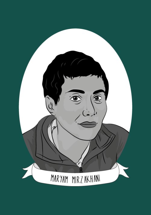
Maryam Mirzakhani was an Iranian mathematician and a professor of mathematics at Stanford University. She was the first-ever female winner of the prestigious Fields Medal prize and the first Iranian to be honoured with the award.
Mirzakhani was born in Tehran, Iran. She attended Farzanegan School, which was part of the National Organization for Development of Exceptional Talents. In both 1994 and 1995 she won the International Mathematical Olympiads for high-school students. In the 1995 International Mathematical Olympiad, she became the first Iranian student to achieve a perfect score and to win two gold medals.
Mirzakhani continued her education at Sharif University of Technology in Tehran, where she earned a BSc in Mathematics. After this, she undertook a a Ph.D. from Harvard University. She worked under the supervision of the Fields Medalist Curtis T. McMullen, and her dissertation focused on Simple Geodesics on Hyperbolic Surfaces and Volume of the Moduli Space of Curves. She had a unique way of working, and “would spend hours on the floor with supersized canvases of paper, sketching out ideas, drawing diagrams and formulae, often leading Anahita [her daughter] to say, “Oh, Mommy is painting again!” Mirzankhani said that “I don’t have any particular recipe [for developing new proofs] … It is like being lost in a jungle and trying to use all the knowledge that you can gather to come up with some new tricks, and with some luck you might find a way out.”
From 2004 to 2008 she was a Clay Mathematics Institute Research Fellow and an assistant professor at Princeton University. She then became a professor at Stanford University where she specialized in theoretical mathematics including moduli spaces, Teichmüller theory, hyperbolic geometry, Ergodic theory and symplectic geometry.”
In 2014, Mirzakhani was awarded the Fields Medal prize for her work on complex geometry and dynamic systems, becoming the first-ever female winner and the first Iranian to be honoured with the award. During her lifetime, she won a number of awards including the 2009 Blumenthal Award for the Advancement of Research in Pure Mathematics and the 2013 Satter Prize of the American Mathematical Society. She worked up until her death in 2017, and was still producing amazing mathematics during her battle with cancer over the last few years.
Sources here, here, here, here and here
The race to map the human body — one cell at a time
The first time molecular biologist Greg Hannon flew through a tumour, he was astonished — and inspired. Using a virtual-reality model, Hannon and his colleagues at the University of Cambridge, UK, flew in and out of blood vessels, took stock of infiltrating immune cells and hatched an idea for an unprecedented tumour atlas.
“Holy crap!” he recalls thinking. “This is going to be just amazing.”
On 10 February, the London-based charity Cancer Research UK announced that Hannon’s team of molecular biologists, astronomers and game designers would receive up to £20 million (US$25 million) over the next five years to develop its interactive virtual-reality map of breast cancers. The tumour that Hannon flew through was a mock-up, but the real models will include data on the expression of thousands of genes and dozens of proteins in each cell of a tumour. The hope is that this spatial and functional detail could reveal more about the factors that influence a tumour’s response to treatment.
The project is just one of a string that aims to build a new generation of cell atlases: maps of organs or tumours that describe location and make-up of each cell in painstaking detail.


![WATCH: Crystal Birth, A Beautiful Timelapse Of Metallic Crystals Forming In Chemical Solutions [video]](https://64.media.tumblr.com/a6b789bd547bb0fc1e3119df87332f1a/tumblr_oghrhqfMiX1rte5gyo2_500.gif)
![WATCH: Crystal Birth, A Beautiful Timelapse Of Metallic Crystals Forming In Chemical Solutions [video]](https://64.media.tumblr.com/0c08f977f51ba218837b72a94f7bbd69/tumblr_oghrhqfMiX1rte5gyo4_500.gif)
![WATCH: Crystal Birth, A Beautiful Timelapse Of Metallic Crystals Forming In Chemical Solutions [video]](https://64.media.tumblr.com/94f32be0db445db0cdfe2546b015295b/tumblr_oghrhqfMiX1rte5gyo1_500.gif)
![WATCH: Crystal Birth, A Beautiful Timelapse Of Metallic Crystals Forming In Chemical Solutions [video]](https://64.media.tumblr.com/333f1f1da60d5ff8500aa7a751dba6c4/tumblr_oghrhqfMiX1rte5gyo3_500.gif)
WATCH: Crystal Birth, a Beautiful Timelapse of Metallic Crystals Forming in Chemical Solutions [video]

Autonomic nervous system
Structure and Function of the Sympathetic and Parasympathetic nervous system
The main function of the autonomic nervous system (ANS) is to assist the body in maintaining a relatively constant internal environment. For example, a sudden increase in systemic blood pressure activates the baroreceptors (those are receptors that detect physical pressure) which in turn modify the activity of the ANS so that the blood pressure is restored to its previous level [1].
The ANS is often regarded as a part of the motor system and is responsible for involuntary action and its effector organs are smooth muscle, cardiac muscle and glands. Another system, the somatic (meaning around the body) nervous system, is responsible for voluntary action in which skeletal muscle is the effector.
The ANS can further be divided into 3 parts: sympathetic, parasympathetic and enteric nervous systems [1][2], with the enteric nervous system sometimes being considered a separate entity [2]. Both parasympathetic and sympathetic nervous systems coexist and work in opposition with each other, ultimately maintaining the correct balance; the activity of one being more active depending on the situation. In a normal resting human, the parasympathetic nervous system dominates, while in a tense and stressful situation, the sympathetic nervous system switches to become dominant.

Figure 1. Structure and function of the central nervous system
This article will be focused on sympathetic and parasympathetic activity from the perspective of:
Anatomy
Biochemical
The sympathetic division provides your “fight or flight” whereas the parasympathetic division helps you to “rest and digest”
Anatomy
Higher centers that control autonomic function include the pons, medulla oblongata and hypothalamus [3].
The pons contains the micturition (urination) and respiratory center.
The medulla oblongata contains the respiratory, cardiac, vomiting, vasomotor and vasodilator centres [4].
The hypothalamus contains the highest concentration of autonomic centres [4]. It contains several centres that control autonomic activities, including heat loss, heat production and conservation, feeding and satiety, as well as fluid intake [4].

Figure 2. Locations of the autonomic control centres of the brain
All 3 structures receive input from certain sources by stimulation of nerve fibres resulting from chemical changes in blood composition like blood pH, blood glucose level, blood osmolarity and volume [4]. Notably, the hypothalamus receives input from cerebral cortex and the limbic system, a system that helps control emotional behaviour [3].
Autonomic promoter neurons are neurons that are found in the brain stem, hypothalamus or even cerebral hemispheres that project to preganglionic neurons (discussed below), where they form synapses with these neurons (5). Hence, input from the higher centres can be relayed to the motor neurons (preganglionic and then postganglionic neurons) which subsequently innervate different body tissues. Changes in the input from these centres could result in responses in those tissues.
The primary functional unit of the sympathetic and parasympathetic nervous system consists of a 2 neuron motor pathway (Figure 3), containing a preganglionic and postganglionic neurons, arranged in series.(2) The two synapse in peripheral ganglion. This clearly distinguishes autonomic motor nervous system and somatic nervous system. The somatic nervous system project from the CNS directly to innervated tissue without any intervening ganglia.(6)

Figure 3. Diagram showing the primary functional unit of the ANS
Sympathetic nervous system
Sympathetic preganglionic neurons mainly are concentrated in the lateral horn in the thoracic (T1-12) and upper lumbar (L1 &2) segments of the spinal cord (Figure 4).
The preganglionic axons leave the spinal cord in 3 ways:
Through the paravertebral ganglion
The preganglionic axon may synapse with postganglionic neurons in this ganglion or some axon may travel rostrally or caudally within the sympathetic trunk before forming synapse with a postganglionic neurons in a different paravertebral ganglion.
Through the prevertebral ganglion
Some preganglionic axons pass the paravertebral ganglion (no synapse occur) and form synapse with postganglionic neurons in prevertebral ganglion, also known as collateral ganglion.
Directly to the organs without any synapse
Some preganglionic axons pass through the sympathetic trunk (no synapse) and end directly on cells of the adrenal medulla, which are equivalent to postganglionic cell.
Parasympathetic nervous system
The parasympathetic preganglionic neurons are located in several cranial nerve nuclei in the brain stem and some are found in the S3 and S4 segments of the sacral spinal cord (Figure 4). The parasympathetic postganglionic neurons are located in cranial ganglia, including the ciliary ganglion, the pterygopalatine, submandibular ganglia, and the otic ganglion. Other ganglia are present near or in the walls of visceral organs. Similarly, the preganglionic neurons form synapse with the postganglionic neurons in the ganglia.

Figure 4. Anatomy of the ANS and how its nuerons innervate tissues
After knowing how nerves connect from the CNS to PNS and to different organs, we will now consider some of the neurotransmitters that are being released at different nerve terminals. It is the binding of these neurotransmitters to the receptors on the effectors that leads to biochemical and physiological changes. Some of the neurotransmitters in use are:
For the synapse between pre and postganglionic neurons mentioned above, the neurotransmitter that is released by the preganglionic axon terminal, is acetylcholine. The corresponding receptors are found on the postsynaptic membrane of postganglionic nerves and are nicotinic receptors.
Parasympathetic postganglionic nerve terminals also release acetylcholine.
Sympathetic postganglionic nerve terminals release mostly noradrenaline
The adrenal medulla receives direct stimulation from sympathetic preganglionic innervation, releases mainly adrenaline (80%) and some noradrenaline into the blood stream. In this case, both adrenaline and noradrenaline act as hormones as they are transported via blood circulating system to target organs instead of neuronal pathway.
Strangely, for the sympathetic postganglionic nerves that innervate the sweat glands, the nerves release acetylcholine (normally only by parasympathetic postganglionic nerve) instead.
1. H.P.Rang, J.M.Ritter, R.J.Flower GH. RANG & DALE’S Pharmacology. In: 8th ed. ELSEVIER CHURCHILL LIVINGSTONE; 2016. p. 145.
2. Bruce M. Koeppen BAS. BERNE & LEVY PHYSIOLOGY. In: 6th ed. MOSBY ELSEVIER; 2010. p. 218.
3. Cholinergic transmission [Internet]. 2015. Available from: http://www.dartmouth.edu/~rpsmith/Cholinergic_Transmission.html
4. Bruce M. Koeppen BAS. BERNE & LEVY PHYSIOLOGY. In: 6th ed. MOSBY ELSEVIER; 2016. p. 44.
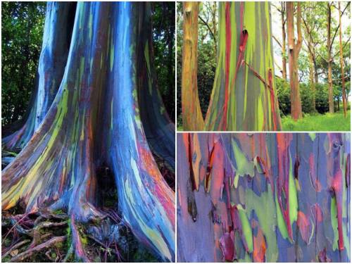
These are rainbow eucalyptus trees (Eucalyptus deglupta) and hail from the Philippine Islands.
The trees get their name from the striking colours observed on their trunks and limbs. Although it may look like someone took a paintbrush to them, these colours are entirely natural. Unlike most trees, the rainbow eucalyptus does not have a thick, cork-like layer of bark on its trunk. The bark is smooth and as it grows it ‘exfoliates’ layers of spent tissue. This exfoliation technique occurs at different stages and in different zones of the tree.
Keep reading
Organic Chemistry, Part 2/6
Reaction Mechanisms: Electrophilic addition to double bonds, SN2, SN1, E1, E2, and the decision tree


Next week: EAS, NAS, pericyclic reactions, Claisen rearrangements, and radical reactions!
Part 1


Cosmos: A Spacetime Odyssey | Super/hypernova + Colors.
-
 debtterbsypcenlli liked this · 1 year ago
 -
 neoncharm8 reblogged this · 3 years ago
 -
 exhausted-pigeon liked this · 4 years ago
 -
 mafia-do-arco-iris reblogged this · 5 years ago
 -
 huge-tromboner reblogged this · 5 years ago
 -
huge-tromboner liked this · 5 years ago
-
 simloey liked this · 5 years ago
 -
 blahblah157 liked this · 5 years ago
 -
 crookedlyfreshtimetravelmattelbo liked this · 5 years ago
 -
 melon-gene liked this · 5 years ago
 -
 wordplay-is-foreplay reblogged this · 5 years ago
 -
 omegalv liked this · 5 years ago
 -
 dr-macadamia-and-his-giraffe liked this · 5 years ago
 -
 deadcrunchyarm liked this · 5 years ago
 -
charswoods liked this · 5 years ago
-
 sinverbauch liked this · 5 years ago
 -
 aldoandco liked this · 5 years ago
 -
 thegaydisneytech reblogged this · 5 years ago
 -
 darllng reblogged this · 5 years ago
 -
darllng liked this · 5 years ago
-
 mayorisus21 reblogged this · 5 years ago
 -
mondocaneus reblogged this · 5 years ago
-
 sj-thefan liked this · 5 years ago
 -
weirdosaround liked this · 5 years ago
-
 flwrmoon liked this · 5 years ago
 -
 milesawayfrmhome reblogged this · 5 years ago
 -
 hobiuwus liked this · 5 years ago
 -
 wilddgirll reblogged this · 5 years ago
 -
wilddgirll liked this · 5 years ago
-
 human-rughts reblogged this · 5 years ago
 -
 raine-y-days liked this · 5 years ago
 -
 quiksilvear reblogged this · 5 years ago
 -
quiksilvear liked this · 5 years ago
-
 elengo4it liked this · 5 years ago
 -
 februarysong1986 reblogged this · 5 years ago
 -
februarysong1986 liked this · 5 years ago
-
 imasoftgay liked this · 5 years ago
A pharmacist and a little science sideblog. "Knowledge belongs to humanity, and is the torch which illuminates the world." - Louis Pasteur
215 posts