Tag Later - Blog Posts


photos of sakurajima, the most active volcano in japan, by takehito miyatake and martin rietze. volcanic storms can rival the intensity of massive supercell thunderstorms, but the source of the charge responsible for this phenomenon remains hotly debated.
in the kind of storm clouds that generate conventional lightning, ice particles and soft hail collide, building up positive and negative charges, respectively. they separate into layers, and the charge builds up until the electric field is high enough to trigger lightning.
but the specific mechanism by which particles of differing charges are separated in the ash cloud is still unknown. lightning has been observed between the eruption plume and the volcano right at the start of an eruption, suggesting that there are processes that occur inside the volcano to lead to charge separation.
volcanic lightning could yield clues about the earth’s geological past, and could answer questions about the beginning of life on our planet. volcanic lightning could have been the essential spark that converted water, hydrogen, ammonia, and methane molecules present on a primeval earth into amino acids, the building blocks of life.
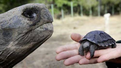
Baby Tortoises Show Up In The Galapagos For The First Time In Over A Century
There hadn’t been one single baby tortoise sighting in more than a century on the Galapagos Island of Pinzon, until a small group of the tiny, shelled youngsters were spotted this year.
The recent births are helping to pull the critically endangered animals back from the brink of extinction after they were nearly laid to waste as a result of human activity.
This is huge news for a species that has been struggling to survive for a century, relying on humans raising young tortoises bred in captivity until they are large enough to not fall prey to rats and predators.


What makes fireworks colorful?
It’s all thanks to the luminescence of metals. When certain metals are heated (over a flame or in a hot explosion) their electrons jump up to a higher energy state. When those electrons fall back down, they emit specific frequencies of light - and each chemical has a unique emission spectrum.
You can see that the most prominent bands in the spectra above match the firework colors. The colors often burn brighter with the addition of an electron donor like Chlorine (Cl).
But the metals alone wouldn’t look like much. They need to be excited. Black powder (mostly nitrates like KNO3) provides oxygen for the rapid reduction of charcoal © to create a lot hot expanding gas - the BOOM. That, in turn, provides the energy for luminescence - the AWWWW.
Aluminium has a special role — it emits a bright white light … and makes sparks!
Images: Charles D. Winters, Andrew Lambert Photography / Science Source, iStockphoto, Epic Fireworks, Softyx, Mark Schellhase, Walkerma, Firetwister, Rob Lavinsky, iRocks.com, Søren Wedel Nielsen

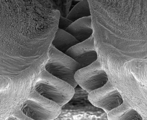
Close-Up of the First Mechanical Gear Ever Found in Nature
The biological form of a mechanical gear was observed in nature for the first time in juvenile planthoppers (Genus: Issus), a common insect that can be found in gardens across Europe.
The insect has hind-leg joints with curved cog-like strips of opposing ‘teeth’ that intermesh, rotating like mechanical gears to synchronize the animal’s legs when it launches into a jump. The finding demonstrates that gear mechanisms previously thought to be solely man-made have an evolutionary precedent.
(Continue Reading)


It’s #InternationalKissingDay! Here’s some topical lipstick chemistry. More info/high-res image: http://wp.me/s4aPLT-lipstick

As Ars has reported previously, scientists have found that triclosan and other antimicrobial soaps have little benefit to consumers and may actually pose risks. These include bolstering antibiotic resistant microbes, giving opportunistic pathogens a leg up, and disrupting microbiomes. In its final ruling, issued Friday, the FDA seemed to agree. “Consumers may think antibacterial washes are more effective at preventing the spread of germs, but we have no scientific evidence that they are any better than plain soap and water,” Janet Woodcock, director of the FDA’s Center for Drug Evaluation and Research (CDER), said in a statement. “In fact, some data suggests that antibacterial ingredients may do more harm than good over the long-term.”
[…]
The ruling does not affect alcohol-based hand sanitizers or wipes, which the agency is reviewing separately. It also does not affect antiseptic products used in healthcare settings.
Asteroid Terms: Explained
There are interesting asteroid characters in our solar system, including an asteroid that has its own moon and even one that is shaped like a dog bone! Our OSIRIS-REx mission launches at 7:05 p.m. EDT today and will travel to asteroid Bennu.

Scientists chose Bennu as the target of the OSIRIS-REx mission because of its composition, size and proximity to Earth. Bennu is a rare B-type asteroid (primitive and carbon-rich), which is expected to have organic compounds and water-bearing minerals like clays.
Our OSIRIS-REx mission will travel to Bennu and bring a small sample back to Earth for study.

When talking about asteroids, there are some terms scientists use that might not be in your typical vocabulary…but we’ll help with that!
Here are a few terms you should know:
Orbital Eccentricity: This number describes the shape of an asteroid’s orbit by how elliptical it is. For asteroids in orbit around the sun, eccentricity is a number between 0 and 1, with 0 being a perfectly circular orbit and 0.99 being a highly elliptical orbit.
Inclination: The angle, in degrees, of how tilted an asteroid’s orbit is compared to another plane of reference, usually the plane of the Earth’s orbit around the sun.
Orbital Period: The number of days it takes for an asteroid to revolve once around the sun. For example, the Earth’s orbital period is 365 days.
Perihelion Distance: The distance between an asteroid and the sun when the asteroid is closest to the sun.
Aphelion Distance: The distance between the asteroid and the sun when the asteroid is farthest away from the sun.
Astronomical unit: A distance unit commonly used to describe orbits of objects around the sun. The distance from the Earth to the sun is one astronomical unit, or 1 AU, equivalent to about 93 million miles or 150 million kilometers.
Diameter: A measure of the size of an asteroid. It is the length of a line from a point on the surface, through the center of the asteroid, extending out to the opposite surface. Irregularly shaped asteroids may have different diameters depending on which direction they are measured.
Rotation Period: The time it takes for an asteroid to complete one revolution around its axis of rotation. For example, the rotation period of the Earth is approximately 24 hours, or 1 day.
Spectral Type: The classification of an asteroid, based on a measurement of the light reflected by the asteroid.

Watch live launch coverage of OSIRIS-REx to asteroid Bennu starting at 5:30 p.m, on NASA TV: http://www.nasa.gov/nasatv
Make sure to follow us on Tumblr for your regular dose of space: http://nasa.tumblr.com


A human brain has around 86 billion neurons, and the communication between these neurons are constant. The sheer scale of these interactions mean a computer (an EEG) can register this electrical activity, with different frequencies indicating different mental states.
Sources
New technique captures the activity of an entire brain in a snapshot
When it comes to measuring brain activity, scientists have tools that can take a precise look at a small slice of the brain (less than one cubic millimeter), or a blurred look at a larger area. Now, researchers at The Rockefeller University have described a new technique that combines the best of both worlds—it captures a detailed snapshot of global activity in the mouse brain.

(Image caption: Sniff, sniff: This density map of the cerebral cortex of a mouse shows which neurons get activated when the animal explores a new environment. The lit up region at the center (white and yellow) represents neurons associated with the mouse’s whiskers)
“We wanted to develop a technique that would show you the level of activity at the precision of a single neuron, but at the scale of the whole brain,” says study author Nicolas Renier, a postdoctoral fellow in the lab of Marc Tessier-Lavigne, Carson Family Professor and head of the Laboratory of Brain Development and Repair, and president of Rockefeller University.
The new method, described in Cell, takes a picture of all the active neurons in the brain at a specific time. The mouse brain contains dozens of millions of neurons, and a typical image depicts the activity of approximately one million neurons, says Tessier-Lavigne. “The purpose of the technique is to accelerate our understanding of how the brain works.”
Making brains transparent
“Because of the nature of our technique, we cannot visualize live brain activity over time—we only see neurons that are active at the specific time we took the snapshot,” says Eliza Adams, a graduate student in Tessier-Lavigne’s lab and co-author of the study. “But what we gain in this trade-off is a comprehensive view of most neurons in the brain, and the ability to compare these active neuronal populations between snapshots in a robust and unbiased manner.”
Here’s how the tool works: The researchers expose a mouse to a situation that would provoke altered brain activity—such as taking an anti-psychotic drug, brushing whiskers against an object while exploring, and parenting a pup—then make the measurement after a pause. The pause is important, explains Renier, because the technique measures neuron activity indirectly, via the translation of neuronal genes into proteins, which takes about 30 minutes to occur.
The researchers then treat the brain to make it transparent—following an improved version of a protocol called iDISCO, developed by Zhuhao Wu, a postdoctoral associate in the Tessier-Lavigne lab—and visualize it using light-sheet microscopy, which takes the snapshot of all active neurons in 3-D.
To determine where an active neuron is located within the brain, Christoph Kirst, a fellow in Rockefeller’s Center for Studies in Physics and Biology, developed software to detect the active neurons and to automatically map the snapshot to a 3-D atlas of the mouse brain, generated by the Allen Brain Institute.
Although each snapshot of brain activity typically includes about one million active neurons, researchers can sift through that mass of data relatively quickly if they compare one snapshot to another snapshot, says Renier. By eliminating the neurons that are active in both images, researchers are left only those specific to each one, enabling them to home in on what is unique to each state.
Observing and testing how the brain works
The primary purpose of the tool, he adds, is to help researchers generate hypotheses about how the brain functions that then can be tested in other experiments. For instance, using their new techniques, the researchers, in collaboration with Catherine Dulac and other scientists at Harvard University, observed that when an adult mouse encounters a pup, a region of its brain known to be active during parenting—called the medial pre-optic nucleus, or MPO—lights up. But they also observed that, after the MPO area becomes activated, there is less activity in the cortical amygdala, an area that processes aversive responses, which they found to be directly connected to the MPO “parenting region.”
“Our hypothesis,” says Renier, “is that parenting neurons put the brake on activity in the fear region, which may suppress aversive responses the mice may have towards pups.” Indeed, mice that are being aggressive to pups tend to show more activity in the cortical amygdala.
To test this idea, the next step is to block the activity of this brain region to see if this reduces aggression in the mice, says Renier.
The technique also has broader implications than simply looking at what areas of the mouse brain are active in different situations, he adds. It could be used to map brain activity in response to any biological change, such as the spread of a drug or disease, or even to explore how the brain makes decisions. “You can use the same strategy to map anything you want in the mouse brain,” says Renier.

Babies don’t just look cute, scientists find
What is it about the sight of an infant that makes almost everyone crack a smile? Big eyes, chubby cheeks, and a button nose? An infectious laugh, soft skin, and a captivating smell? While we have long known that babies look cute, Oxford University researchers have found that cuteness is designed to appeal to all our senses.
They explain that all these characteristics contribute to ‘cuteness’ and trigger our caregiving behaviours, which is vital because infants need our constant attention to survive and thrive. The study is published in the journal Trends in Cognitive Sciences.
Morten Kringelbach, who together with Eloise Stark, Catherine Alexander, Professor Marc Bornstein and Professor Alan Stein, led the work in the Department of Psychiatry at the University of Oxford, said: ‘Infants attract us through all our senses, which helps make cuteness one of the most basic and powerful forces shaping our behaviour.’
Reviewing the emerging literature on how cute infants and animals affect the brain, the Oxford University team found that cuteness supports key parental capacities by igniting fast privileged neural activity followed by slower processing in large brain networks also involved in play, empathy, and perhaps even higher-order moral emotions.
The data shows that definitions of cuteness should not be limited just to visual features but include positive infant sounds and smells. From an evolutionary standpoint, cuteness is a very potent protective mechanism that ensures survival for otherwise completely dependent infants.
Professor Kringelbach said: ‘This is the first evidence of its kind to show that cuteness helps infants to survive by eliciting caregiving, which cannot be reduced to simple, instinctual behaviours. Instead, caregiving involves a complex choreography of slow, careful, deliberate, and long-lasting prosocial behaviours, which ignite fundamental brain pleasure systems that are also engaged when eating food or listening to music, and always involve pleasant experiences.’
The study shows that cuteness affects both men and women, even those without children.
‘This might be a fundamental response present in everyone, regardless of parental status or gender, and we are currently conducting the first long-term study of what happens to brain responses when we become parents.’ said Kringelbach.

i’m proud of them

From retina to cortex: An unexpected division of labor
Neurons in our brain do a remarkable job of translating sensory information into reliable representations of our world that are critical to effectively guide our behavior. The parts of the brain that are responsible for vision have long been center stage for scientists’ efforts to understand the rules that neural circuits use to encode sensory information. Years of research have led to a fairly detailed picture of the initial steps of this visual process, carried out in the retina, and how information from this stage is transmitted to the visual part of the cerebral cortex, a thin sheet of neurons that forms the outer surface of the brain. We have also learned much about the way that neurons represent visual information in visual cortex, as well as how different this representation is from the information initially supplied by the retina. Scientists are now working to understand the set of rules—the neural blueprint— that explains how these representations of visual information in the visual cortex are constructed from the information provided by the retina. Using the latest functional imaging techniques, scientists at MPFI have recently discovered a surprisingly simple rule that explains how neural circuits combine information supplied by different types of cells in the retina to build a coherent, information-rich representation of our visual world.
Vision begins with the spatial pattern of light and dark that falls on the retinal surface. One important function performed by the neural circuits in the visual cortex is the preservation of the orderly spatial relationships of light versus dark that exist on the retinal surface. These neural circuits form an orderly map of visual space where each point on the surface of the cortex contains a column of neurons that each respond to a small region of visual space— and adjacent columns respond to adjacent regions of visual space. But these cortical circuits do more than build a map of visual space: individual neurons within these columns each respond selectively to the specific orientation of edges in their region of visual space; some neurons respond preferentially to vertical edges, some to horizontal edges, and others to angles in between. This property is also mapped in a columnar fashion where all neurons in a radial column have the same orientation preference, and adjacent columns prefer slightly different orientations.
Things would be easy if all the cortex had to do was build a map of visual space: a simple one to one mapping of points on the retinal surface to columns in the cortex would be all that was necessary. But building a map of orientation that coexists with the map of visual space is a much greater challenge. This is because the neurons of the retina do not distinguish orientation in the first step of vision. Instead, information on the orientation of edges must be constructed by neural circuits in the visual cortex. This is done using information supplied from two distinct types of retinal cells: those that respond to increases in light (ON-cells) and those that respond to decreases in light (OFF-cells). Adding to the complexity, orientation selectivity depends on having individual cortical neurons receive their ON and OFF signals from non-overlapping regions of visual space, and the spatial arrangement of these regions determines the orientation preference of the cell. Cortical neurons that prefer vertical edge orientations have ON and OFF responsive regions that are displaced horizontally in visual space, those that prefer horizontal edge orientations have their ON and OFF regions displaced vertically in visual space, and this systematic relationship holds for all other edge orientations.
So cortical circuits face a paradox: How do they take the spatial information from the retina and distort it to create an orderly map of orientation selectivity, while at the same time preserving fine retinal spatial information in order to generate an orderly map of visual space? Nature’s solution might best be called ‘divide and conquer’. By using imaging technologies that allow visualization of the ON and OFF response regions of hundreds of individual cortical neurons, Kuo-Sheng Lee and Sharon Huang in David Fitzpatrick’s lab at MPFI have discovered that fine scale retinal spatial information is preserved by the OFF response regions of cortical neurons, while the ON response regions exhibit systematic spatial displacements that are necessary to build an orderly map of edge orientation. Preserving the detailed spatial information from the retina in the OFF response regions is consistent with evidence that dark elements of natural scenes convey more fine scale information than the light elements, and that OFF retinal neurons have properties that allow them to better extract this information. In addition, Lee et al. show that this OFF-anchored cortical architecture enables emergence of an additional orderly map of absolute spatial phase—a property that hasn’t received much attention from neuroscientists, but computer vision research has shown contains a wealth of information about the visual scene that can be used to efficiently encode spatial patterns, motion, and depth.
While these are important new insights into how visual information is transformed from retina to cortical representations, they pose a host of new questions about the network of synaptic connections that performs this transformation, and the developmental mechanisms that construct it, questions that the Fitzpatrick Lab continues to explore.

It turns out that there’s actually a scientific reason behind why people don’t sleep soundly in an unfamiliar place.
According to a study conducted by Brown University, the first night that you sleep in a new environment the left hemisphere of the brain stays alert while the right hemisphere rests. The biological trait is thought to have allowed early humans to respond to nighttime threats. “When we’re sleeping in a new environment and we don’t know how many predators are around, it would make sense to keep half the brain more alert and more responsive to bumps in the night,” said Niels Rattenborg, who led the study.
The evolutionary advantage this brain traits provides is probably less useful to modern humans — making us experience less restful sleep when we spend the night at a hotel or friend’s house, for example — but in the animal kingdom, this trait still helps marine mammals such as dolphins, whales, and seals respond to threats during their slumber.


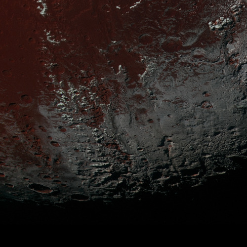
This is one slice of an incredible high resolution, enhanced color image of Pluto, recently released by NASA. You can see the full, larger version here.
Credit: NASA/JHUAPL/SwRI
The Hubble Space telescope just sent back a new photo of the Twin Jet Nebula. Here’s what it looked like in 1997:

And now …

Whoa. But wait, we also got an updated image of the merging galaxies NGC 6240. What it looked like in 2008:

And today:

Science, you’re the best. Oh, and the explanation behind those merging galaxies and their black holes is wild.

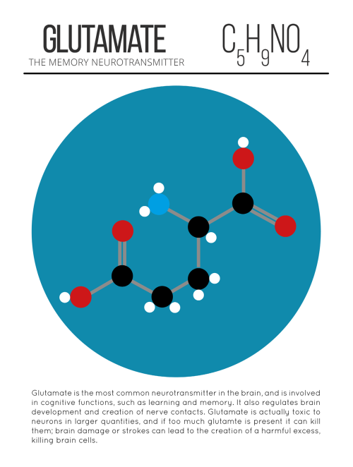
Glutamate, an essential food for the brain
Glutamate is an amino acid with very different functions: in the pancreas, it modulates the activity of the pancreatic ß-cells responsible for insulin production, whereas in the brain it is the main excitatory neurotransmitter. In recent years, it has been suspected to play an additional role in the functioning of the brain. By discovering how the brain uses glutamate to produce energy, researchers at the University of Geneva (UNIGE) confirm this hypothesis and highlight unexpected links with the rest of the body. To read in Cell Reports.
Unlike other organs, the brain cannot draw its energy from lipids, an energy resource widely present in the body. The blood-brain barrier, which protects it from the pathogens and toxins circulating in the blood, indeed limits the passage of these lipids. Moreover, while most of the organs in the human body have the ability to store glucose by increasing their mass, the brain, prisoner of the cranial bones, cannot count on these variations in volume. Unable to store its food, it depends on sugar supplied in real-time by the rest of the body. This distribution of energy is controlled by the liver.
Pierre Maechler, professor at the Faculty of Medicine at UNIGE, and his team therefore decided to verify if glutamate was indeed an energy source for the brain. To do so, the researchers analyzed the role of the glutamate dehydrogenase enzyme in the brain. In mutant form, this enzyme, encoded by the Glud1 gene, is responsible for a congenital hyperinsulinism syndrome, a severe disease affecting at the same time the endocrine pancreas, the liver and the brain. Individuals affected by this syndrome suffer from intellectual disability and have a high risk of epilepsy. “We have suppressed the Glud1 gene in the brain of mice. In the absence of glutamate dehydrogenase, we observed that the brain was no longer able to convert glutamate into energy, even though the amino acid was present in the brain,” explains Melis Karaca, first author of this study.
Priority to the brain
Devoid of the energy supplied by cerebral glutamate, the brain sends signals to the liver to requisition a compensatory proportion of glucose, at the expense of the rest of the body. This is why the transgenic mice also showed a growth deficit and muscle atrophy. “This clearly shows how the brain works in a just-in-time manner and that each percent of energy resources is essential for its proper functioning,” highlights Professor Pierre Maechler. “If a part of this energy disappears, the brain serves itself first and the rest of the body suffers. The liver must then make more glucose by drawing upon muscle protein, resulting in loss of muscle mass. Knowing that the brain uses glutamate as an energy resource allows us to reflect on other ways to overcome a potential shortfall. ”
Scientists also suspect a correlation between the Glud1 gene and some neurodevelopmental disorders, particularly epilepsy and schizophrenia. They are currently pursuing their research by introducing in mice the same Glud1 mutation detected in epileptic patients. At the same time, another group is working with schizophrenic patients to assess the way their brain uses glutamate.


Missed any of the graphics featured in C&EN? They’ve now put a page together so you can find all of the graphics in one place, on subjects including Guinness, daffodils, barbecue & more: ow.ly/RB10e
which scientist should you fight
geologist: will throw copious amounts of rocks at you. not recommended unless you can also throw equal amounts of rocks back
botanist: knows 1001 ways to poison you. probably shouldn't fight
zoologist: knows 1001 animals that can kill you. probably shouldn't fight either
entomologist: spiders. enough said.
physiologist: they know too much about the human body and how to cause optimal pain with minimal damage. not safe.
geneticist: will unleash their army of mutated fruit flies at you. can be either good or bad thing, depending on your preference for flies with legs growing out of their eyes
immunologist: they have perfected the t-cell inspired technique of "death by neglect". if you fight them you will die in the saddest way possible
microbiologist: please don't fight someone who is already pissed about antibiotic resistance and can identify bacteria based solely on their smell
climatologist: will choose the battlefield as somewhere in the path of a category 5 hurricane and then leave you to die. do not fight please
environmental scientist: they can control the entire world do you really want to fight them
chemist: have you seen breaking bad? no, do not fight them. do NOT
physicist: will kill you with math. not the best way to go
herpetologist: can probably speak parseltongue and know just which frogs are best at taking over your habitat. only fight if you live in antarctica
cancer biologist: has immediate access to at least 5 different tumor cell lines and knows exactly where to inject them in your heart to cause metastases. don't even look them in the eye
marine biologist: is a real life aquaman. will lure you with cute river otters and then finish you off with some terrifying deep sea creature. better to just stay home and never leave
psychologist: is basically a mind reader. will drown you in your deepest darkest fears. 10/10 do not recommend to fight
molecular biologist: will kill you organelle by organelle. you will die a slow and painful death while covered in budding yeast
statistician: their power is always over 80%, and they will quickly punt you in the path of a normal distribution even before you can yell "Wilcoxon!"
archaeologist: can use a trowel 59 different ways, and only 9 are for digging. one can only guess the other 50, so may be advisable to stay far away
astronomer: will launch you into space and send you to a planet so inhospitable not even matt damon can make it back this time
pharmacologist: why would you ever fight someone who knows all about drugs. why
computer scientist: they know the perfect algorithm for death. do not fight, even with a firewall
linguist: no matter where you are, they can talk about you behind your back in the native tongue. do you really want death by humiliation. do you
dinosaurologist: are you kidding me?? the answer is no
sociologist: yea

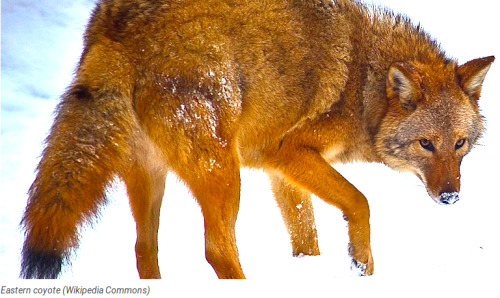
A new species is evolving before scientists’ eyes in the eastern United States.
Wolves faced with a diminishing number of potential mates are lowering their standards and mating with other, similar species, reported The Economist.
The interbreeding began up to 200 years ago, as European settlers pushed into southern Ontario and cleared the animal’s habitat for farming and killed a large number of the wolves that lived there.
That also allowed coyotes to spread from the prairies, and the white farmers brought dogs into the region.
Over time, wolves began mating with their new, genetically similar neighbors.
The resulting offspring — which has been called the eastern coyote or, to some, the “coywolf” — now number in the millions, according to researchers at North Carolina State University.
Interspecies-bred animals are typically less vigorous than their parents, The Economist reported — if the offspring survive at all.
That’s not the case at all with the wolf-coyote-dog hybrid, which has developed into a sum greater than the whole of its parts.
At about 55 pounds, the hybrid animal is about twice as heavy as a standard coyote, and her large jaws, faster legs and muscular body allow her to take down small deer and even hunt moose in packs, and the animal is skilled at hunting in both open terrain and dense woodland.
An analysis of 437 hybrid animals found that coyote DNA dominates her genetic makeup, with about one-tenth of its DNA from dogs, usually larger dogs such as Doberman pinschers and German shepherds, and a quarter from wolves.
The animal’s cry starts out as a deep-pitched wolf howl that morphs into higher-pitched yipping — like a coyote.
Her dog DNA may carry an additional advantage.
Some scientists think the hybrid animal is able to adapt to city life — which neither coyotes or wolves have managed to do on their own — because her dog ancestry allows her to tolerate people and noise.
The coywolves have spread into some of the nation’s largest cities — including New York, Boston and Washington — using railway corridors.
The interbreeding allows the animal to diversify her diet and eat discarded food, along with rodents and smaller mammals — including cats, which coywolves eat skull and all — and they have evolved to become nocturnal to avoid humans.
The animals are also smart enough to learn to look both ways before crossing roads.
Not all researchers agree the animal is a distinct species, arguing that one species does not interbreed with another — although the hybrid’s existence raises the question of whether wolves and coyotes are distinct species in the first place.
But scientists who have studied the animal say the mixing of genes has been much faster, extensive and transformational than anyone had noticed until fairly recently.
“(This) amazing contemporary evolution story (is) happening right underneath our nose,” said Roland Kays, a researcher at North Carolina State.
Watch this report on coywolves.
Raw Story


Olinguito
On Tuesday, Bill Stanley grabbed my arm and pulled me into a side hallway as we were walking towards the mammal collections on the third floor. He looked around suspiciously before leaning in, and in a hushed tone he said
there’s a new raccoon.
What do you mean?
There’s a new raccoon. You can’t tell anyone. It’s in the pipeline. Going live on Thursday.
Wha- I wasn’t going to-
You can’t tell anyone. Guess where it was discovered?
Oh, geez. I don’t know. Maybe Per-
Here. It was discovered here.
Then he patted my shoulder, winked, and kept walking.
Such was my introduction to the olinguito - and yesterday Bill brought it out to show me. In front of us were two drawers, one with the previously known species and the newly described animals on the right. It was immediately obvious to me that before us were two different animals - the size, color and length of the fur, the size of the ears - but without the previous knowledge that they were not one in the same, would I have seen the same dissimilarities?
This is what sparks me. This is what drives my enthusiasm. In these drawers for sixty years, side-by-side these animals remained, their full potential not realized until a curious researcher took the quiet time to sit down and take a concentrated look at them. It’s the romance of the discovery – it was here all along! – and once you see the striking inconsistencies there comes a feeling of empowerment, the thought that we are the next big discoverers. The thrill of the breakthrough remains attainable, accessible. It’s not beyond our reach or out of grasp - I look forward to seeing what you find next.

Both hemispheres of the brain process numbers
Researchers of the Jena University (Germany) and of the Jena University Hospital located an important region for the visual processing of numbers in the human brain and showed that it is active in both hemispheres. In the ’Journal of Neuroscience’ the scientists published high resolution magnetic resonance recordings of this region.
The human brain works with division of labour. Although our thinking organ excels in displaying amazing flexibility and plasticity, typically different areas of the brain take over different tasks. While words and language are mainly being processed in the left hemisphere, the right hemisphere is responsible for numerical reasoning. According to previous findings, this division of labour originates from the fact that the first steps in the processing of letters and numbers are also located individually in the different hemispheres. But this is not the case, at least not when it comes to the visual processing of numbers.
Neuroscientists of the Friedrich Schiller University Jena and of the Jena University Hospital discovered that the visual processing of numbers takes place in a so-called ‘visual number form area’ (NFA) - in fact in both hemispheres alike. The Jena scientists were the first to publish high resolution magnetic resonance recordings showing the activity in this region of the brain of healthy test persons. The area is normally difficult to get access to.
The 'blind spot’ in the brain
In their study Dr. Mareike Grotheer and Prof. Dr. Gyula Kovács from the Institute for Psychology of Jena University as well as Dr. Karl-Heinz Herrmann from the Department of Radiology (IDIR) of the Jena University Hospital presented subjects with numbers, letters and pictures of everyday objects. Meanwhile the participants’ brain activity was recorded using magnetic resonance imaging (MRI). Thus the researchers were able to clearly identify the region in which the visual processing of numbers takes place. The small area at the underside of the left and right temporal lobe reacted with increased activity at the presentation of numbers. Letters and other images but also false numbers lead to a significantly lower brain activity in this area.
Although the Jena team already knew from other scientists’ previous research where they had to look for the area, a lot of developmental work went into the newly published story. “This region has been a kind of blind spot in the human brain until now,” Mareike Grotheer says. And here is why: Hidden underneath the ear and the acoustic meatus, surrounded by bone and air, previous MRI scans showed a number of artefacts and thus obstructed detailed research.
For their study the Jena scientists used a high-performance 3 tesla MRI scanner of the Institute of Diagnostic and Interventional Radiology (IDIR) of the Jena University Hospital. They recorded three-dimensional images of the brain of the test subjects at an unusually high spatial resolution and hence with only very few artefacts. In addition these recordings were spatially smoothed whereby the remaining 'white noise’ could be removed. This approach will help other scientists to investigate a part of the brain that until now had been nearly inaccessible. “In this region not only numbers are being processed but also faces and objects,” Prof. Kovács states.